1GAI

GLUCOAMYLASE-471 COMPLEXED WITH D-GLUCO-DIHYDROACARBOSE


Experimental Data Snapshot

  • Method: X-RAY DIFFRACTION
  • Resolution: 1.70 Å
  • R-Value Free: 0.167 
  • R-Value Work: 0.147 
  • R-Value Observed: 0.147 

wwPDB Validation   3D Report Full Report


Ligand Structure Quality Assessment 


This is version 2.1 of the entry. See complete history


Literature

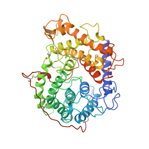
Crystallographic complexes of glucoamylase with maltooligosaccharide analogs: relationship of stereochemical distortions at the nonreducing end to the catalytic mechanism.

Aleshin, A.E.Stoffer, B.Firsov, L.M.Svensson, B.Honzatko, R.B.

(1996) Biochemistry 35: 8319-8328

  • DOI: https://doi.org/10.1021/bi960321g
  • Primary Citation of Related Structures:  
    1GAH, 1GAI

  • PubMed Abstract: 

    Crystal structures at pH 4 of complexes of glucoamylase from Aspergillus awamori var. X100 with the pseudotetrasaccharides D-gluco-dihydroacarbose and acarbose have been refined to R-factors of 0.147 and 0.131 against data to 1.7- and 2.0-A resolution, respectively. The two inhibitors bind in nearly identical manners, each exhibiting a dual binding mode with respect to the location of the last sugar residues. The reduced affinity of D-gluco-dihydroacarbose (K1 = 10(-8) M) relative to acarbose (K1 = 10(-12) M) may stem in part from the weakening of hydrogen bonds of the catalytic water (Wat 500) to the enzyme. Steric contacts between the nonreducing end of D-gluco-dihydroacarbose and the catalytic water perturb Wat 500 from its site of optimal hydrogen bonding to the active site. Interactions within the active site displace the 6-hydroxymethyl group of the nonreducing end of both acarbose and D-gluco-dihydroacarbose toward a more axial position. In the case of D-gluco-dihydroacarbose the shift in the position of the 6-hydroxymethyl group occurs with a 12 degrees change in two dihedral angles of the glucopyranose ring toward a half-chair conformation. The observed conformational distortion of the first residue of D-gluco-dihydroacarbose is consistent with the generation of a glucopyranosyl cation in the transition state. Comparable distortions of stereochemistry in model compounds require approximately 2 kcal/mol, not more than 25% of the energy necessary to form the half-chair conformation in glucose. The magnitude of stereochemical distortion observed in the active site of glucoamylase suggests that favorable electrostatic interactions between the putative glucopyranosyl cation intermediate and the active site must be more important in stabilizing the transition state than mechanical distortion of the substrate.


  • Organizational Affiliation

    Department of Biochemistry and Biophysics, Iowa State University, Ames, 50011, USA.


Macromolecules
Find similar proteins by:  (by identity cutoff)  |  3D Structure
Entity ID: 1
MoleculeChains Sequence LengthOrganismDetailsImage
GLUCOAMYLASE-471472Aspergillus awamoriMutation(s): 0 
EC: 3.2.1.3
UniProt
Find proteins for P69327 (Aspergillus awamori)
Explore P69327 
Go to UniProtKB:  P69327
Entity Groups  
Sequence Clusters30% Identity50% Identity70% Identity90% Identity95% Identity100% Identity
UniProt GroupP69327
Sequence Annotations
Expand
  • Reference Sequence
Oligosaccharides

Help

Entity ID: 2
MoleculeChains Length2D Diagram Glycosylation3D Interactions
alpha-D-mannopyranose-(1-2)-alpha-D-mannopyranose-(1-3)-beta-D-mannopyranose-(1-4)-2-acetamido-2-deoxy-beta-D-glucopyranose-(1-4)-2-acetamido-2-deoxy-beta-D-glucopyranose
B
5N-Glycosylation
Glycosylation Resources
GlyTouCan:  G42227JK
GlyCosmos:  G42227JK
GlyGen:  G42227JK
Entity ID: 3
MoleculeChains Length2D Diagram Glycosylation3D Interactions
alpha-D-mannopyranose-(1-2)-alpha-D-mannopyranose-(1-2)-alpha-D-mannopyranose-(1-3)-[alpha-D-mannopyranose-(1-3)-[alpha-D-mannopyranose-(1-6)]alpha-D-mannopyranose-(1-6)]beta-D-mannopyranose-(1-4)-2-acetamido-2-deoxy-beta-D-glucopyranose-(1-4)-2-acetamido-2-deoxy-beta-D-glucopyranose
C
9N-Glycosylation
Glycosylation Resources
GlyTouCan:  G68668TB
GlyCosmos:  G68668TB
GlyGen:  G68668TB
Entity ID: 4
MoleculeChains Length2D Diagram Glycosylation3D Interactions
4,6-dideoxy-4-{[(1S,2S,3S,4R,5R)-2,3,4-trihydroxy-5-(hydroxymethyl)cyclohexyl]amino}-alpha-D-glucopyranose-(1-4)-alpha-D-glucopyranose-(1-4)-alpha-D-glucopyranose
D
3N/AN/A
Glycosylation Resources
GlyTouCan:  G55244EK
GlyCosmos:  G55244EK
Biologically Interesting Molecules (External Reference) 1 Unique
Experimental Data & Validation

Experimental Data

  • Method: X-RAY DIFFRACTION
  • Resolution: 1.70 Å
  • R-Value Free: 0.167 
  • R-Value Work: 0.147 
  • R-Value Observed: 0.147 
  • Space Group: P 21 21 21
Unit Cell:
Length ( Å )Angle ( ˚ )
a = 116.9α = 90
b = 104.4β = 90
c = 48.4γ = 90
Software Package:
Software NamePurpose
XENGENdata collection
XENGENdata reduction
X-PLORmodel building
X-PLORrefinement
XENGENdata scaling
X-PLORphasing

Structure Validation

View Full Validation Report


Ligand Structure Quality Assessment 


Entry History 

Deposition Data

Revision History  (Full details and data files)

  • Version 1.0: 1996-08-17
    Type: Initial release
  • Version 1.1: 2008-03-10
    Changes: Version format compliance
  • Version 1.2: 2011-07-13
    Changes: Atomic model, Version format compliance
  • Version 1.3: 2011-10-05
    Changes: Derived calculations
  • Version 2.0: 2020-07-29
    Type: Remediation
    Reason: Carbohydrate remediation
    Changes: Advisory, Atomic model, Data collection, Database references, Derived calculations, Non-polymer description, Other, Structure summary
  • Version 2.1: 2024-04-03
    Changes: Data collection, Database references, Refinement description, Structure summary