1F11

F124 FAB FRAGMENT FROM A MONOCLONAL ANTI-PRES2 ANTIBODY


Experimental Data Snapshot

  • Method: X-RAY DIFFRACTION
  • Resolution: 3.00 Å
  • R-Value Free: 0.271 
  • R-Value Work: 0.184 
  • R-Value Observed: 0.191 

wwPDB Validation   3D Report Full Report


This is version 1.4 of the entry. See complete history


Literature

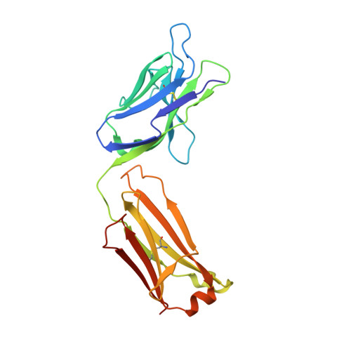
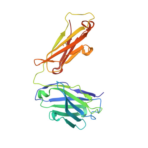
Structure of the Fab fragment from F124, a monoclonal antibody specific for hepatitis B surface antigen.

Saul, F.A.Vulliez-Le Normand, B.Passafiume, M.Riottot, M.M.Bentley, G.A.

(2000) Acta Crystallogr D Biol Crystallogr 56: 945-951

  • DOI: https://doi.org/10.1107/s0907444900008088
  • Primary Citation of Related Structures:  
    1F11

  • PubMed Abstract: 

    The crystal structure of the Fab fragment from the monoclonal anti-preS2 antibody F124 (IgG1,kappa) has been solved by molecular replacement and refined at 3.0 A resolution. The Fab crystallizes with two independent molecules in the asymmetric unit. F124 recognizes an epitope contained within the preS2 segment between residues 120 and 132 of the surface antigen of hepatitis B virus. The antibody shows a high affinity for the glycan N-linked to Asn123, but it also cross-reacts with the non-glycosylated peptide fragment 120-132. Although crystallization was performed in the presence of an eightfold excess of the cross-reactive peptide, no evidence for the ligand was found in the antigen-binding site, which is close to a neighbouring molecule in the crystal lattice. The antigen-binding site has a groove-like topology which is modulated with pocket-like cavities. It is characterized by a large number of tyrosine and aspartate residues. The importance of germ-line mutations at the binding site is discussed.


  • Organizational Affiliation

    Unité d'Immunologie Structurale, CNRS URA 2185, Institut Pasteur, 25 Rue du Dr Roux, 75724 Paris, France.


Macromolecules
Find similar proteins by:  (by identity cutoff)  |  3D Structure
Entity ID: 1
MoleculeChains Sequence LengthOrganismDetailsImage
F124 IMMUNOGLOBULIN (KAPPA LIGHT CHAIN)
A, C
218Mus musculusMutation(s): 0 
UniProt
Find proteins for P01837 (Mus musculus)
Explore P01837 
Go to UniProtKB:  P01837
Find proteins for P01665 (Mus musculus)
Explore P01665 
Go to UniProtKB:  P01665
Entity Groups  
Sequence Clusters30% Identity50% Identity70% Identity90% Identity95% Identity100% Identity
UniProt GroupsP01665P01837
Sequence Annotations
Expand
  • Reference Sequence
Find similar proteins by:  (by identity cutoff)  |  3D Structure
Entity ID: 2
MoleculeChains Sequence LengthOrganismDetailsImage
F124 IMMUNOGLOBULIN (IGG1 HEAVY CHAIN)
B, D
221Mus musculusMutation(s): 0 
UniProt
Find proteins for Q9D8L4 (Mus musculus)
Explore Q9D8L4 
Go to UniProtKB:  Q9D8L4
Entity Groups  
Sequence Clusters30% Identity50% Identity70% Identity90% Identity95% Identity100% Identity
UniProt GroupQ9D8L4
Sequence Annotations
Expand
  • Reference Sequence
Experimental Data & Validation

Experimental Data

  • Method: X-RAY DIFFRACTION
  • Resolution: 3.00 Å
  • R-Value Free: 0.271 
  • R-Value Work: 0.184 
  • R-Value Observed: 0.191 
  • Space Group: C 1 2 1
Unit Cell:
Length ( Å )Angle ( ˚ )
a = 135.7α = 90
b = 54β = 101
c = 120γ = 90
Software Package:
Software NamePurpose
DENZOdata reduction
SCALEPACKdata scaling
AMoREphasing
X-PLORrefinement

Structure Validation

View Full Validation Report


Entry History 

Deposition Data

Revision History  (Full details and data files)

  • Version 1.0: 2000-08-30
    Type: Initial release
  • Version 1.1: 2008-04-27
    Changes: Version format compliance
  • Version 1.2: 2011-07-13
    Changes: Version format compliance
  • Version 1.3: 2017-06-21
    Changes: Database references, Source and taxonomy, Structure summary
  • Version 1.4: 2023-08-09
    Changes: Advisory, Data collection, Database references, Refinement description