Structural insight into the binding diversity between the Tyr-phosphorylated human ephrinBs and Nck2 SH2 domain.
Ran, X., Song, J.(2005) J Biological Chem 280: 19205-19212
- PubMed: 15764601
- DOI: https://doi.org/10.1074/jbc.M500330200
- Primary Citation of Related Structures:
1Z3K - PubMed Abstract:
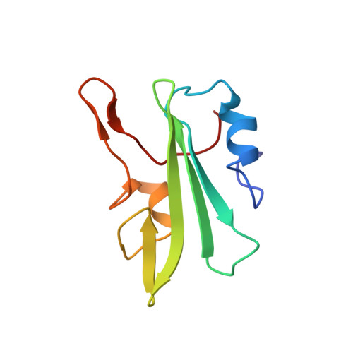
The binding interaction between the Nck2 SH2 domain and the phosphorylated ephrinB initiates a critical pathway for the reverse signaling network mediated by Eph receptor-ephrinB. Previously, the NMR structure and Tyr phosphorylations of the human ephrinB cytoplasmic domain have been studied. To obtain a complete story, it would be of significant interest to determine the structure of the Nck2 SH2 domain that shows a low sequence identity to other SH2 domains with known structures. Here, we report the determination of the solution structure of the human Nck2 SH2 domain and investigate its interactions with three phosphorylated ephrinB fragments by NMR spectroscopy. The results indicate that: 1) although the human Nck2 SH2 domain adopts a core tertiary fold common to all SH2 domains, it owns some unique properties such as a shorter C-terminal helix and unusual electrostatic potential surface. However, the most striking finding is that the C-terminal tail of the human Nck2 SH2 domain adopts a short antiparallel beta-sheet that, to the best of our knowledge, has never been identified in other SH2 domains. The truncation study suggests that one function of the C-terminal tail is to control the folding/solubility of the SH2 domain. 2) In addition to [Tyr(P)304]ephrinB2(301-322) and [Tyr(P)316]ephrinB2(301-322), here we identified [Tyr(P)330]ephrinB2(324-333) also capable of binding to the SH2 domain. The detailed NMR study indicated that the binding mechanisms for the three ephrinB fragments might be different. The binding with [Tyr(P)304]-ephrinB2(301-322) and [Tyr(P)316]ephrinB2(301-322) might be mostly involved in the residues over the N-half of the SH2 domain and provoked a significant increase in the backbone and side chain dynamics of the SH2 domain on the microsecond-millisecond time scale. In contrast, binding with [Tyr(P)330]ephrinB2(324-333) might have most residues over both halves engaged but induced less profound conformational dynamics on the mus-ms time scale.
- Department of Biochemistry, National University of Singapore, 10 Kent Ridge Crescent, Singapore 119260.
Organizational Affiliation: