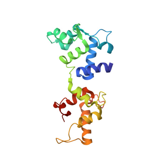
The crystal structure of calcium- and integrin-binding protein 1: Insights into redox regulated functions
Blamey, C.J., Ceccarelli, C., Naik, U.P., Bahnson, B.J.(2005) Protein Sci 14: 1214-1221
- PubMed: 15840829
- DOI: https://doi.org/10.1110/ps.041270805
- Primary Citation of Related Structures:
1Y1A - PubMed Abstract:
Calcium- and integrin-binding protein 1 (CIB1) is involved in the process of platelet aggregation by binding the cytoplasmic tail of the alpha(IIb) subunit of the platelet-specific integrin alpha(Iib)beta(3). Although poorly understood, it is widely believed that CIB1 acts as a global signaling regulator because it is expressed in many tissues that do not express integrin alpha(Iib)beta(3). We report the structure of human CIB1 to a resolution of 2.3 A, crystallized as a dimer. The dimer interface includes an extensive hydrophobic patch in a crystal form with 80% solvent content. Although the dimer form of CIB1 may not be physiologically relevant, this intersub-unit surface is likely to be linked to alpha(IIb) binding and to the binding of other signaling partner proteins. The C-terminal domain of CIB1 is structurally similar to other EF-hand proteins such as calmodulin and calcineurin B. Despite structural homology to the C-terminal domain, the N-terminal domain of CIB1 lacks calcium-binding sites. The structure of CIB1 revealed a complex with a molecule of glutathione in the reduced state bond to the N-terminal domain of one of the two subunits poised to interact with the free thiol of C35. Glutathione bound in this fashion suggests CIB1 may be redox regulated. Next to the bound GSH, the orientation of residues C35, H31, and S48 is suggestive of a cysteine-type protein phosphatase active site. The potential enzymatic activity of CIB1 is discussed and suggests a mechanism by which it regulates a wide variety of proteins in cells in addition to platelets.
- Department of Biological Sciences, University of Delaware, Newark, DE 19716, USA.
Organizational Affiliation: