Optimal isotope labelling for NMR protein structure determinations.
Kainosho, M., Torizawa, T., Iwashita, Y., Terauchi, T., Mei Ono, A., Guntert, P.(2006) Nature 440: 52-57
- PubMed: 16511487
- DOI: https://doi.org/10.1038/nature04525
- Primary Citation of Related Structures:
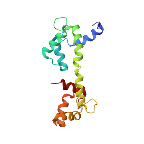
1X02, 2D21 - PubMed Abstract:
Nuclear-magnetic-resonance spectroscopy can determine the three-dimensional structure of proteins in solution. However, its potential has been limited by the difficulty of interpreting NMR spectra in the presence of broadened and overlapping resonance lines and low signal-to-noise ratios. Here we present stereo-array isotope labelling (SAIL), a technique that can overcome many of these problems by applying a complete stereospecific and regiospecific pattern of stable isotopes that is optimal with regard to the quality and information content of the resulting NMR spectra. SAIL uses exclusively chemically and enzymatically synthesized amino acids for cell-free protein expression. We demonstrate for the 17-kDa protein calmodulin and the 41-kDa maltodextrin-binding protein that SAIL offers sharpened lines, spectral simplification without loss of information, and the ability to rapidly collect the structural restraints required to solve a high-quality solution structure for proteins twice as large as commonly solved by NMR. It thus makes a large class of proteins newly accessible to detailed solution structure determination.
- CREST/JST and Graduate School of Science, Tokyo Metropolitan University, 1-1 Minami-ohsawa, Hachioji, 192-0397, Japan. kainosho@nmr.chem.metro-u.ac.jp
Organizational Affiliation: