


crystal structure of JW1657 from Escherichia coli
Kato-Murayama, M., Shirouzu, M., Yokoyama, S.To be published.
Experimental Data Snapshot
wwPDB Validation 3D Report Full Report

Entity ID: 1 | |||||
|---|---|---|---|---|---|
| Molecule | Chains | Sequence Length | Organism | Details | Image |
| Protein ydhR | 123 | Escherichia coli | Mutation(s): 2 EC: 1 | 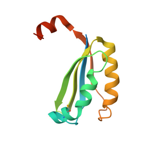 | |
UniProt | |||||
Find proteins for P0ACX3 (Escherichia coli (strain K12)) Explore P0ACX3 Go to UniProtKB: P0ACX3 | |||||
Entity Groups | |||||
| Sequence Clusters | 30% Identity50% Identity70% Identity90% Identity95% Identity100% Identity | ||||
| UniProt Group | P0ACX3 | ||||
Sequence AnnotationsExpand | |||||
| |||||
| Modified Residues 1 Unique | |||||
|---|---|---|---|---|---|
| ID | Chains | Type | Formula | 2D Diagram | Parent |
| MSE Query on MSE | A, B | L-PEPTIDE LINKING | C5 H11 N O2 Se |  | MET |
| Length ( Å ) | Angle ( ˚ ) |
|---|---|
| a = 85.803 | α = 90 |
| b = 85.803 | β = 90 |
| c = 95.923 | γ = 120 |
| Software Name | Purpose |
|---|---|
| CNS | refinement |
| HKL-2000 | data reduction |
| SCALEPACK | data scaling |
| SOLVE | phasing |


