


Crystal Structure of Uroporphyrin-III-C-Methyltrans from Thermus Thermophilus
Jeyakanthan, J., Tahirov, T.H.To be published.
Experimental Data Snapshot
wwPDB Validation 3D Report Full Report

Entity ID: 1 | |||||
|---|---|---|---|---|---|
| Molecule | Chains | Sequence Length | Organism | Details | Image |
| Uroporphyrin-III C-methyltransferase | 235 | Thermus thermophilus | Mutation(s): 0 EC: 2.1.1.107 | 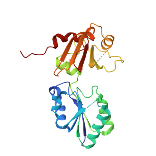 | |
UniProt | |||||
Find proteins for Q53WA2 (Thermus thermophilus (strain ATCC 27634 / DSM 579 / HB8)) Explore Q53WA2 Go to UniProtKB: Q53WA2 | |||||
Entity Groups | |||||
| Sequence Clusters | 30% Identity50% Identity70% Identity90% Identity95% Identity100% Identity | ||||
| UniProt Group | Q53WA2 | ||||
Sequence AnnotationsExpand | |||||
| |||||
| Length ( Å ) | Angle ( ˚ ) |
|---|---|
| a = 54.609 | α = 90 |
| b = 87.924 | β = 90 |
| c = 91.602 | γ = 90 |
| Software Name | Purpose |
|---|---|
| CNS | refinement |
| HKL-2000 | data reduction |
| SCALEPACK | data scaling |
| SOLVE | phasing |


