1TVM

NMR structure of enzyme GatB of the galactitol-specific phosphoenolpyruvate-dependent phosphotransferase system


Experimental Data Snapshot

  • Method: SOLUTION NMR
  • Conformers Calculated: 100 
  • Conformers Submitted: 20 
  • Selection Criteria: structures with the least restraint violations, structures with the lowest energy 

wwPDB Validation   3D Report Full Report


This is version 1.3 of the entry. See complete history


Literature

NMR structure of the enzyme GatB of the galactitol-specific phosphoenolpyruvate-dependent phosphotransferase system and its interaction with GatA.

Volpon, L.Young, C.R.Matte, A.Gehring, K.

(2006) Protein Sci 15: 2435-2441

  • DOI: https://doi.org/10.1110/ps.062337406
  • Primary Citation of Related Structures:  
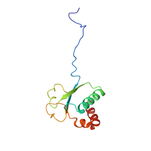
    1TVM

  • PubMed Abstract: 

    The phosphoenolpyruvate-dependent carbohydrate transport system (PTS) couples uptake with phosphorylation of a variety of carbohydrates in prokaryotes. In this multienzyme complex, the enzyme II (EII), a carbohydrate-specific permease, is constituted of two cytoplasmic domains, IIA and IIB, and a transmembrane channel IIC domain. Among the five families of EIIs identified in Escherichia coli, the galactitol-specific transporter (II(gat)) belongs to the glucitol family and is structurally the least well-characterized. Here, we used nuclear magnetic resonance (NMR) spectroscopy to solve the three-dimensional structure of the IIB subunit (GatB). GatB consists of a central four-stranded parallel beta-sheet flanked by alpha-helices on both sides; the active site cysteine of GatB is located at the beginning of an unstructured loop between beta1 and alpha1 that folds into a P-loop-like structure. This structural arrangement shows similarities with other IIB subunits but also with mammalian low molecular weight protein tyrosine phosphatases (LMW PTPase) and arsenate reductase (ArsC). An NMR titration was performed to identify the GatA-interacting residues.


  • Organizational Affiliation

    Department of Biochemistry, McGill University, Montreal, Quebec H3G 1Y6, Canada. laurent.volpon@umontreal.ca


Macromolecules
Find similar proteins by:  (by identity cutoff)  |  3D Structure
Entity ID: 1
MoleculeChains Sequence LengthOrganismDetailsImage
PTS system, galactitol-specific IIB component113Escherichia coliMutation(s): 0 
Gene Names: GATBC2618Z3256ECS2896SF2155S2281
EC: 2.7.1.69
UniProt
Find proteins for P0A435 (Escherichia coli O6:H1 (strain CFT073 / ATCC 700928 / UPEC))
Explore P0A435 
Go to UniProtKB:  P0A435
Entity Groups  
Sequence Clusters30% Identity50% Identity70% Identity90% Identity95% Identity100% Identity
UniProt GroupP0A435
Sequence Annotations
Expand
  • Reference Sequence
Experimental Data & Validation

Experimental Data

  • Method: SOLUTION NMR
  • Conformers Calculated: 100 
  • Conformers Submitted: 20 
  • Selection Criteria: structures with the least restraint violations, structures with the lowest energy 

Structure Validation

View Full Validation Report


Entry History 

Revision History  (Full details and data files)

  • Version 1.0: 2005-09-06
    Type: Initial release
  • Version 1.1: 2008-04-30
    Changes: Version format compliance
  • Version 1.2: 2011-07-13
    Changes: Version format compliance
  • Version 1.3: 2022-03-02
    Changes: Data collection, Database references, Derived calculations