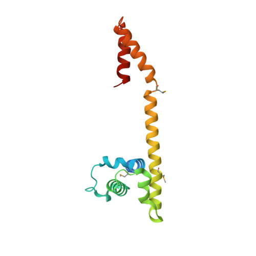
1T3W

Crystal Structure of the E.coli DnaG C-terminal domain (residues 434 to 581)


Experimental Data Snapshot

  • Method: X-RAY DIFFRACTION
  • Resolution: 2.80 Å
  • R-Value Free: 0.308 
  • R-Value Work: 0.269 
  • R-Value Observed: 0.271 

wwPDB Validation   3D Report Full Report


This is version 1.3 of the entry. See complete history


Literature

Crystal and solution structures of the helicase-binding domain of Escherichia coli primase

Oakley, A.J.Loscha, K.V.Schaeffer, P.M.Liepinsh, E.Pintacuda, G.Wilce, M.C.J.Otting, G.Dixon, N.E.

(2005) J Biol Chem 280: 11495-11504

  • DOI: https://doi.org/10.1074/jbc.M412645200
  • Primary Citation of Related Structures:  
    1T3W

  • PubMed Abstract: 

    During bacterial DNA replication, the DnaG primase interacts with the hexameric DnaB helicase to synthesize RNA primers for extension by DNA polymerase. In Escherichia coli, this occurs by transient interaction of primase with the helicase. Here we demonstrate directly by surface plasmon resonance that the C-terminal domain of primase is responsible for interaction with DnaB6. Determination of the 2.8-angstroms crystal structure of the C-terminal domain of primase revealed an asymmetric dimer. The monomers have an N-terminal helix bundle similar to the N-terminal domain of DnaB, followed by a long helix that connects to a C-terminal helix hairpin. The connecting helix is interrupted differently in the two monomers. Solution studies using NMR showed that an equilibrium exists between a monomeric species with an intact, extended but naked, connecting helix and a dimer in which this helix is interrupted in the same way as in one of the crystal conformers. The other conformer is not significantly populated in solution, and its presence in the crystal is due largely to crystal packing forces. It is proposed that the connecting helix contributes necessary structural flexibility in the primase-helicase complex at replication forks.


  • Organizational Affiliation

    Research School of Chemistry, Australian National University, Canberra, Australian Capital Territory 0200, Australia.


Macromolecules
Find similar proteins by:  (by identity cutoff)  |  3D Structure
Entity ID: 1
MoleculeChains Sequence LengthOrganismDetailsImage
DNA primase
A, B
148Escherichia coliMutation(s): 0 
Gene Names: dnaGdnaPparB
EC: 2.7.7
UniProt
Find proteins for P0ABS5 (Escherichia coli (strain K12))
Explore P0ABS5 
Go to UniProtKB:  P0ABS5
Entity Groups  
Sequence Clusters30% Identity50% Identity70% Identity90% Identity95% Identity100% Identity
UniProt GroupP0ABS5
Sequence Annotations
Expand
  • Reference Sequence
Small Molecules
Ligands 1 Unique
IDChains Name / Formula / InChI Key2D Diagram3D Interactions
ACY
Query on ACY

Download Ideal Coordinates CCD File 
C [auth B]ACETIC ACID
C2 H4 O2
QTBSBXVTEAMEQO-UHFFFAOYSA-N
Modified Residues  1 Unique
IDChains TypeFormula2D DiagramParent
MSE
Query on MSE
A, B
L-PEPTIDE LINKINGC5 H11 N O2 SeMET
Experimental Data & Validation

Experimental Data

  • Method: X-RAY DIFFRACTION
  • Resolution: 2.80 Å
  • R-Value Free: 0.308 
  • R-Value Work: 0.269 
  • R-Value Observed: 0.271 
  • Space Group: I 41 2 2
Unit Cell:
Length ( Å )Angle ( ˚ )
a = 142.239α = 90
b = 142.239β = 90
c = 192.131γ = 90
Software Package:
Software NamePurpose
REFMACrefinement
DENZOdata reduction
SCALEPACKdata scaling
CNSphasing

Structure Validation

View Full Validation Report


Entry History 

Deposition Data

Revision History  (Full details and data files)

  • Version 1.0: 2004-11-02
    Type: Initial release
  • Version 1.1: 2008-04-30
    Changes: Version format compliance
  • Version 1.2: 2011-07-13
    Changes: Advisory, Derived calculations, Refinement description, Version format compliance
  • Version 1.3: 2016-09-28
    Changes: Other