


Crystal structure of refined tetragonal crystal of YodA from Escherichia coli
Shin, D.H., Yokota, H., Kim, R., Kim, S.H.To be published.
Experimental Data Snapshot
wwPDB Validation 3D Report Full Report

Entity ID: 1 | |||||
|---|---|---|---|---|---|
| Molecule | Chains | Sequence Length | Organism | Details | Image |
| Metal-binding Protein yodA | 216 | Escherichia coli | Mutation(s): 0 Gene Names: yodA | 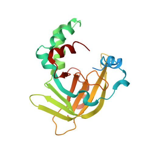 | |
UniProt | |||||
Find proteins for P76344 (Escherichia coli (strain K12)) Explore P76344 Go to UniProtKB: P76344 | |||||
Entity Groups | |||||
| Sequence Clusters | 30% Identity50% Identity70% Identity90% Identity95% Identity100% Identity | ||||
| UniProt Group | P76344 | ||||
Sequence AnnotationsExpand | |||||
| |||||
| Ligands 1 Unique | |||||
|---|---|---|---|---|---|
| ID | Chains | Name / Formula / InChI Key | 2D Diagram | 3D Interactions | |
| ZN Query on ZN | B [auth A], C [auth A], D [auth A], E [auth A] | ZINC ION Zn PTFCDOFLOPIGGS-UHFFFAOYSA-N |  | ||
| Length ( Å ) | Angle ( ˚ ) |
|---|---|
| a = 55.009 | α = 90 |
| b = 55.009 | β = 90 |
| c = 153.785 | γ = 90 |
| Software Name | Purpose |
|---|---|
| CNS | refinement |
| DENZO | data reduction |
| SCALEPACK | data scaling |
| AMoRE | phasing |


