1S0L

Interleukin 1 beta mutant F42W


Experimental Data Snapshot

  • Method: X-RAY DIFFRACTION
  • Resolution: 2.34 Å
  • R-Value Free: 0.222 
  • R-Value Work: 0.191 
  • R-Value Observed: 0.192 

wwPDB Validation   3D Report Full Report


This is version 1.4 of the entry. See complete history


Literature

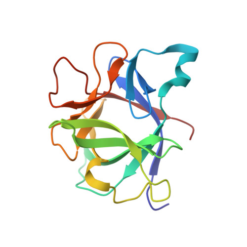
Structural and energetic consequences of mutations in a solvated hydrophobic cavity.

Adamek, D.H.Guerrero, L.Blaber, M.Caspar, D.L.D.

(2005) J Mol Biol 346: 307-318

  • DOI: https://doi.org/10.1016/j.jmb.2004.11.046
  • Primary Citation of Related Structures:  
    1S0L, 1T4Q, 1TOO, 1TP0, 1TWE, 1TWM

  • PubMed Abstract: 

    The structural and energetic consequences of modifications to the hydrophobic cavity of interleukin 1-beta (IL-1beta) are described. Previous reports demonstrated that the entirely hydrophobic cavity of IL-1beta contains positionally disordered water. To gain a better understanding of the nature of this cavity and the water therein, a number of mutant proteins were constructed by site-directed mutagenesis, designed to result in altered hydrophobicity of the cavity. These mutations involve the replacement of specific phenylalanine residues, which circumscribe the cavity, with tyrosine, tryptophan, leucine and isoleucine. Using differential scanning calorimetry to determine the relative stabilities of the wild-type and mutant proteins, we found all of the mutants to be destabilizing. X-ray crystallography was used to identify the structural consequences of the mutations. No clear correlation between the hydrophobicities of the specific side-chains introduced and the resulting stabilities was found.


  • Organizational Affiliation

    Institute of Molecular Biophysics, Florida State University, Tallahassee, FL 32306-4380, USA. daniel.adamek@msfc.nasa.gov


Macromolecules
Find similar proteins by:  (by identity cutoff)  |  3D Structure
Entity ID: 1
MoleculeChains Sequence LengthOrganismDetailsImage
Interleukin-1 beta153Homo sapiensMutation(s): 1 
Gene Names: IL1B
UniProt & NIH Common Fund Data Resources
Find proteins for P01584 (Homo sapiens)
Explore P01584 
Go to UniProtKB:  P01584
PHAROS:  P01584
GTEx:  ENSG00000125538 
Entity Groups  
Sequence Clusters30% Identity50% Identity70% Identity90% Identity95% Identity100% Identity
UniProt GroupP01584
Sequence Annotations
Expand
  • Reference Sequence
Experimental Data & Validation

Experimental Data

  • Method: X-RAY DIFFRACTION
  • Resolution: 2.34 Å
  • R-Value Free: 0.222 
  • R-Value Work: 0.191 
  • R-Value Observed: 0.192 
  • Space Group: P 43
Unit Cell:
Length ( Å )Angle ( ˚ )
a = 54.9α = 90
b = 54.9β = 90
c = 77.24γ = 90
Software Package:
Software NamePurpose
DENZOdata reduction
SCALEPACKdata scaling
CNSrefinement
CNSphasing

Structure Validation

View Full Validation Report


Entry History 

Deposition Data

Revision History  (Full details and data files)

  • Version 1.0: 2004-03-30
    Type: Initial release
  • Version 1.1: 2008-04-29
    Changes: Version format compliance
  • Version 1.2: 2011-07-13
    Changes: Version format compliance
  • Version 1.3: 2021-10-27
    Changes: Database references
  • Version 1.4: 2023-08-23
    Changes: Data collection, Refinement description