1RWJ

c7-type three-heme cytochrome domain


Experimental Data Snapshot

  • Method: X-RAY DIFFRACTION
  • Resolution: 1.70 Å
  • R-Value Free: 0.218 
  • R-Value Work: 0.195 
  • R-Value Observed: 0.195 

wwPDB Validation   3D Report Full Report


Ligand Structure Quality Assessment 


This is version 2.0 of the entry. See complete history


Literature

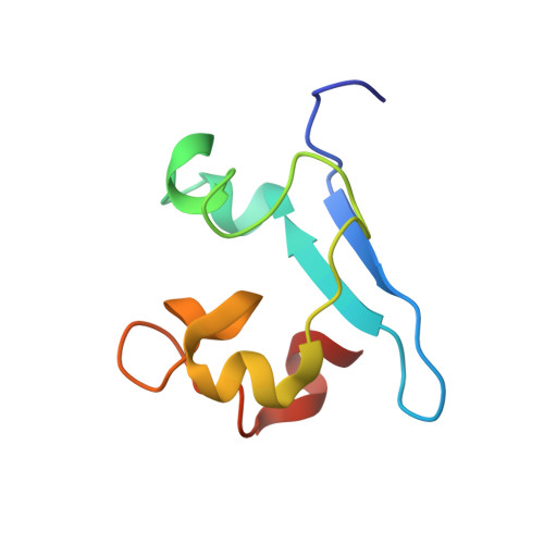
Structure of a novel c7-type three-heme cytochrome domain from a multidomain cytochrome c polymer.

Pokkuluri, P.R.Londer, Y.Y.Duke, N.E.C.Erickson, J.Pessanha, M.Salgueiro, C.A.Schiffer, M.

(2004) Protein Sci 13: 1684-1692

  • DOI: https://doi.org/10.1110/ps.04626204
  • Primary Citation of Related Structures:  
    1RWJ

  • PubMed Abstract: 

    The structure of a novel c(7)-type cytochrome domain that has two bishistidine coordinated hemes and one heme with histidine, methionine coordination (where the sixth ligand is a methionine residue) was determined at 1.7 A resolution. This domain is a representative of domains that form three polymers encoded by the Geobacter sulfurreducens genome. Two of these polymers consist of four and one protein of nine c(7)-type domains with a total of 12 and 27 hemes, respectively. Four individual domains (termed A, B, C, and D) from one such multiheme cytochrome c (ORF03300) were cloned and expressed in Escherichia coli. The domain C produced diffraction quality crystals from 2.4 M sodium malonate (pH 7). The structure was solved by MAD method and refined to an R-factor of 19.5% and R-free of 21.8%. Unlike the two c(7) molecules with known structures, one from G. sulfurreducens (PpcA) and one from Desulfuromonas acetoxidans where all three hemes are bishistidine coordinated, this domain contains a heme which is coordinated by a methionine and a histidine residue. As a result, the corresponding heme could have a higher potential than the other two hemes. The apparent midpoint reduction potential, E(app), of domain C is -105 mV, 50 mV higher than that of PpcA.


  • Organizational Affiliation

    Biosciences Division, Argonne National Laboratory, Argonne, IL 60439, USA.


Macromolecules
Find similar proteins by:  (by identity cutoff)  |  3D Structure
Entity ID: 1
MoleculeChains Sequence LengthOrganismDetailsImage
Cytochrome c family protein82Geobacter sulfurreducensMutation(s): 0 
Gene Names: ORF03300
UniProt
Find proteins for Q74BP5 (Geobacter sulfurreducens (strain ATCC 51573 / DSM 12127 / PCA))
Explore Q74BP5 
Go to UniProtKB:  Q74BP5
Entity Groups  
Sequence Clusters30% Identity50% Identity70% Identity90% Identity95% Identity100% Identity
UniProt GroupQ74BP5
Sequence Annotations
Expand
  • Reference Sequence
Experimental Data & Validation

Experimental Data

  • Method: X-RAY DIFFRACTION
  • Resolution: 1.70 Å
  • R-Value Free: 0.218 
  • R-Value Work: 0.195 
  • R-Value Observed: 0.195 
  • Space Group: P 41 21 2
Unit Cell:
Length ( Å )Angle ( ˚ )
a = 43.8α = 90
b = 43.8β = 90
c = 123.8γ = 90
Software Package:
Software NamePurpose
CNSrefinement
HKL-2000data reduction
SCALEPACKdata scaling
CNSphasing

Structure Validation

View Full Validation Report


Ligand Structure Quality Assessment 


Entry History 

Deposition Data

Revision History  (Full details and data files)

  • Version 1.0: 2004-08-03
    Type: Initial release
  • Version 1.1: 2008-04-29
    Changes: Version format compliance
  • Version 1.2: 2011-07-13
    Changes: Version format compliance
  • Version 2.0: 2021-03-03
    Changes: Advisory, Atomic model, Data collection, Derived calculations, Non-polymer description, Structure summary