1RPE

THE PHAGE 434 OR2/R1-69 COMPLEX AT 2.5 ANGSTROMS RESOLUTION


Experimental Data Snapshot

  • Method: X-RAY DIFFRACTION
  • Resolution: 2.50 Å
  • R-Value Observed: 0.209 

wwPDB Validation   3D Report Full Report


This is version 1.3 of the entry. See complete history


Literature

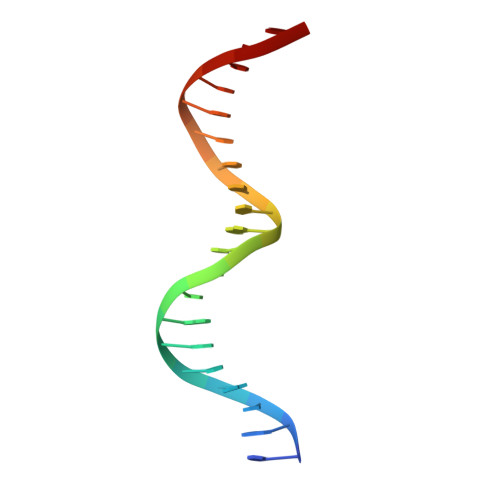
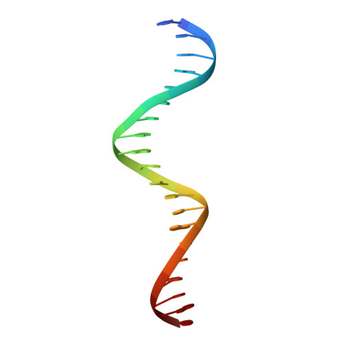
The phage 434 OR2/R1-69 complex at 2.5 A resolution.

Shimon, L.J.Harrison, S.C.

(1993) J Mol Biol 232: 826-838

  • DOI: https://doi.org/10.1006/jmbi.1993.1434
  • Primary Citation of Related Structures:  
    1RPE

  • PubMed Abstract: 

    The crystal structure of the DNA-binding domain of bacteriophage 434 repressor (R1-69) in complex with a 20 base-pair DNA fragment has been determined to 2.5 A resolution. The DNA fragment contains the sequence of the OR2 operator site, which differs from the previously studied OR1 site at three of the variable six central base-pairs. Comparison of the two structures shows that the overall bent conformation of the DNA backbone as well as the pattern of DNA-protein interactions seen in the OR1/R1-69 complex are maintained in the OR2 complex. However, the conformations of the DNA base-pairs are different in the two structures. In particular, the central base-pairs of OR2/R1-69 structure are more co-planar than in OR1/R1-69, and there are no cross-strand "bifurcated" hydrogen bonds. These results show that binding of the protein causes operator DNA to adopt a particular, well-defined backbone conformation, and they reinforce the notion that the energetic cost of achieving this conformation, most likely different for different sequences, can determine, at least in part, the relative affinity of the repressor for different operator sites.


  • Organizational Affiliation

    Howard Hughes Medical Institute, Harvard University Department of Biochemistry and Molecular Biology, Cambridge, MA 02138.


Macromolecules

Find similar proteins by:  (by identity cutoff)  |  3D Structure
Entity ID: 3
MoleculeChains Sequence LengthOrganismDetailsImage
PROTEIN (434 REPRESSOR)C [auth L],
D [auth R]
69Phage 434Mutation(s): 0 
UniProt
Find proteins for P16117 (Enterobacteria phage 434)
Explore P16117 
Go to UniProtKB:  P16117
Entity Groups  
Sequence Clusters30% Identity50% Identity70% Identity90% Identity95% Identity100% Identity
UniProt GroupP16117
Sequence Annotations
Expand
  • Reference Sequence

Find similar nucleic acids by:  Sequence   |   3D Structure  

Entity ID: 1
MoleculeChains LengthOrganismImage
DNA (5'-D(*TP*AP*TP*AP*CP*AP*AP*TP*GP*TP*AP*TP*CP*TP*TP*GP*T P*TP*TP*G)-3')A [auth B]20N/A
Sequence Annotations
Expand
  • Reference Sequence

Find similar nucleic acids by:  Sequence   |   3D Structure  

Entity ID: 2
MoleculeChains LengthOrganismImage
DNA (5'-D(*AP*CP*AP*AP*AP*CP*AP*AP*GP*AP*TP*AP*CP*AP*TP*TP*G P*TP*AP*T)-3')B [auth A]20N/A
Sequence Annotations
Expand
  • Reference Sequence
Experimental Data & Validation

Experimental Data

  • Method: X-RAY DIFFRACTION
  • Resolution: 2.50 Å
  • R-Value Observed: 0.209 
  • Space Group: P 21 21 21
Unit Cell:
Length ( Å )Angle ( ˚ )
a = 149.29α = 90
b = 64.29β = 90
c = 27.69γ = 90
Software Package:
Software NamePurpose
CORELSrefinement

Structure Validation

View Full Validation Report


Entry History 

Deposition Data

Revision History  (Full details and data files)

  • Version 1.0: 1994-01-31
    Type: Initial release
  • Version 1.1: 2008-05-22
    Changes: Version format compliance
  • Version 1.2: 2011-07-13
    Changes: Version format compliance
  • Version 1.3: 2024-02-14
    Changes: Data collection, Database references