Crystal structure of KsgA, a universally conserved rRNA adenine dimethyltransferase in Escherichia coli
O'Farrell, H.C., Scarsdale, J.N., Rife, J.P.(2004) J Mol Biology 339: 337-353
- PubMed: 15136037
- DOI: https://doi.org/10.1016/j.jmb.2004.02.068
- Primary Citation of Related Structures:
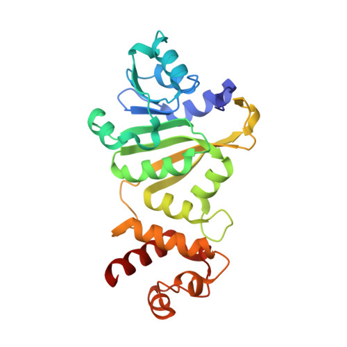
1QYR - PubMed Abstract:
The bacterial enzyme KsgA catalyzes the transfer of a total of four methyl groups from S-adenosyl-l-methionine (S-AdoMet) to two adjacent adenosine bases in 16S rRNA. This enzyme and the resulting modified adenosine bases appear to be conserved in all species of eubacteria, eukaryotes, and archaebacteria, and in eukaryotic organelles. Bacterial resistance to the aminoglycoside antibiotic kasugamycin involves inactivation of KsgA and resulting loss of the dimethylations, with modest consequences to the overall fitness of the organism. In contrast, the yeast ortholog, Dim1, is essential. In yeast, and presumably in other eukaryotes, the enzyme performs a vital role in pre-rRNA processing in addition to its methylating activity. Another ortholog has been discovered recently, h-mtTFB in human mitochondria, which has a second function; this enzyme is a nuclear-encoded mitochondrial transcription factor. The KsgA enzymes are homologous to another family of RNA methyltransferases, the Erm enzymes, which methylate a single adenosine base in 23S rRNA and confer resistance to the MLS-B group of antibiotics. Despite their sequence similarity, the two enzyme families have strikingly different levels of regulation that remain to be elucidated. We have crystallized KsgA from Escherichia coli and solved its structure to a resolution of 2.1A. The structure bears a strong similarity to the crystal structure of ErmC' from Bacillus stearothermophilus and a lesser similarity to sc-mtTFB, the Saccharomyces cerevisiae version of h-mtTFB. Comparison of the three crystal structures and further study of the KsgA protein will provide insight into this interesting group of enzymes.
- Department of Biochemistry, Virginia Commonwealth University, Richmond VA 23298-0133, USA.
Organizational Affiliation: