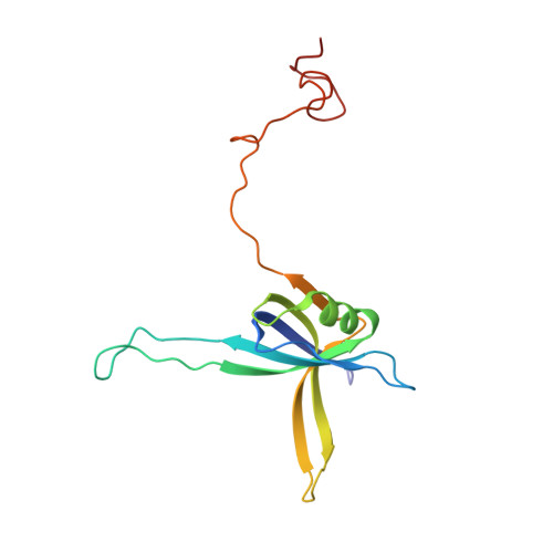
1QVC

CRYSTAL STRUCTURE ANALYSIS OF SINGLE STRANDED DNA BINDING PROTEIN (SSB) FROM E.COLI


Experimental Data Snapshot

  • Method: X-RAY DIFFRACTION
  • Resolution: 2.20 Å
  • R-Value Free: 0.317 
  • R-Value Work: 0.247 
  • R-Value Observed: 0.247 

wwPDB Validation   3D Report Full Report


This is version 1.4 of the entry. See complete history


Literature

Roles of functional loops and the C-terminal segment of a single-stranded DNA binding protein elucidated by X-Ray structure analysis.

Matsumoto, T.Morimoto, Y.Shibata, N.Kinebuchi, T.Shimamoto, N.Tsukihara, T.Yasuoka, N.

(2000) J Biochem 127: 329-335

  • DOI: https://doi.org/10.1093/oxfordjournals.jbchem.a022611
  • Primary Citation of Related Structures:  
    1EQQ, 1QVC

  • PubMed Abstract: 

    The single-stranded DNA (ssDNA) binding protein from Escherichia coli (EcoSSB) plays a central role in DNA replication, recombination and repair. The tertiary structure of EcoSSB was determined at 2.2 A resolution. This is rather higher resolution than previously reported. Crystals were grown from the homogeneous intact protein but the EcoSSB tetramer in the crystals contains truncated subunits lacking a part of the C-terminal. The structure determined includes biologically important flexible loops and C-terminal regions, and revealed the existence of concavities. These concavities include the residues important for ssDNA binding. An ssDNA can be fitted on the concavities and further stabilized through interactions with the loops forming flexible lids. It seems likely to play a central role in the binding of ssDNA.


  • Organizational Affiliation

    Department of Life Science, Himeji Institute of Technology, Kamigori, Ako-gun, Hyogo 678-1297, Japan.


Macromolecules
Find similar proteins by:  (by identity cutoff)  |  3D Structure
Entity ID: 1
MoleculeChains Sequence LengthOrganismDetailsImage
SINGLE STRANDED DNA BINDING PROTEIN MONOMER
A, B, C, D
145Escherichia coliMutation(s): 0 
UniProt
Find proteins for P0AGE0 (Escherichia coli (strain K12))
Explore P0AGE0 
Go to UniProtKB:  P0AGE0
Entity Groups  
Sequence Clusters30% Identity50% Identity70% Identity90% Identity95% Identity100% Identity
UniProt GroupP0AGE0
Sequence Annotations
Expand
  • Reference Sequence
Experimental Data & Validation

Experimental Data

  • Method: X-RAY DIFFRACTION
  • Resolution: 2.20 Å
  • R-Value Free: 0.317 
  • R-Value Work: 0.247 
  • R-Value Observed: 0.247 
  • Space Group: C 1 2 1
Unit Cell:
Length ( Å )Angle ( ˚ )
a = 105.369α = 90
b = 62.923β = 112.57
c = 97.792γ = 90
Software Package:
Software NamePurpose
X-PLORmodel building
CNSrefinement
DENZOdata reduction
SCALEPACKdata scaling
X-PLORphasing

Structure Validation

View Full Validation Report


Entry History 

Deposition Data

Revision History  (Full details and data files)

  • Version 1.0: 2000-06-05
    Type: Initial release
  • Version 1.1: 2008-04-27
    Changes: Version format compliance
  • Version 1.2: 2011-07-13
    Changes: Version format compliance
  • Version 1.3: 2012-07-25
    Changes: Other
  • Version 1.4: 2024-02-14
    Changes: Data collection, Database references