1OXM

STRUCTURE OF CUTINASE


Experimental Data Snapshot

  • Method: X-RAY DIFFRACTION
  • Resolution: 2.30 Å
  • R-Value Free: 0.227 
  • R-Value Work: 0.163 
  • R-Value Observed: 0.163 

wwPDB Validation   3D Report Full Report


Ligand Structure Quality Assessment 


This is version 1.3 of the entry. See complete history


Literature

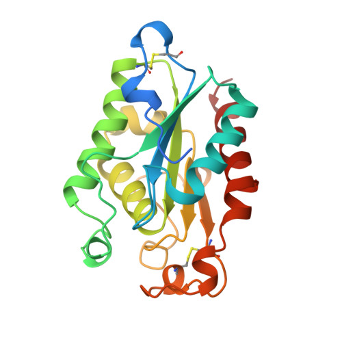
Crystal structure of cutinase covalently inhibited by a triglyceride analogue.

Longhi, S.Mannesse, M.Verheij, H.M.De Haas, G.H.Egmond, M.Knoops-Mouthuy, E.Cambillau, C.

(1997) Protein Sci 6: 275-286

  • DOI: https://doi.org/10.1002/pro.5560060202
  • Primary Citation of Related Structures:  
    1OXM

  • PubMed Abstract: 

    Cutinase from Fusarium solani is a lipolytic enzyme that hydrolyses triglycerides efficiently. All the inhibited forms of lipolytic enzymes described so far are based on the use of small organophosphate and organophosphonate inhibitors, which bear little resemblance to a natural triglyceride substrate. In this article we describe the crystal structure of cutinase covalently inhibited by (R)-1,2-dibutyl-carbamoylglycero-3-O-p-nitrophenylbutyl-phos phonate, a triglyceride analogue mimicking the first tetrahedral intermediate along the reaction pathway. The structure, which has been solved at 2.3 A, reveals that in both the protein molecules of the asymmetric unit the inhibitor is almost completely embedded in the active site crevice. The overall shape of the inhibitor is that of a fork: the two dibutyl-carbamoyl chains point towards the surface of the protein, whereas the butyl chain bound to the phosphorous atom is roughly perpendicular to the sn-1 and sn-2 chains. The sn-3 chain is accommodated in a rather small pocket at the bottom of the active site crevice, thus providing a structural explanation for the preference of cutinase for short acyl chain substrates.


  • Organizational Affiliation

    Architecture et Fonction des Macromolécules Biologiques, UPR 9039, CNRS, Marseille, France.


Macromolecules
Find similar proteins by:  (by identity cutoff)  |  3D Structure
Entity ID: 1
MoleculeChains Sequence LengthOrganismDetailsImage
CUTINASE
A, B
214Fusarium vanetteniiMutation(s): 0 
EC: 3.1.1
Membrane Entity: Yes 
UniProt
Find proteins for P00590 (Fusarium vanettenii)
Explore P00590 
Go to UniProtKB:  P00590
Entity Groups  
Sequence Clusters30% Identity50% Identity70% Identity90% Identity95% Identity100% Identity
UniProt GroupP00590
Sequence Annotations
Expand
  • Reference Sequence
Small Molecules
Ligands 1 Unique
IDChains Name / Formula / InChI Key2D Diagram3D Interactions
TC4
Query on TC4

Download Ideal Coordinates CCD File 
C [auth A],
D [auth B]
BUTYL-PHOSPHINIC ACID 2,3-BIS-BUTYLCARBAMOYLOXY-PROPYL ESTER GROUP
C17 H35 N2 O6 P
QWIXWBNJNKHHHW-OAHLLOKOSA-N
Experimental Data & Validation

Experimental Data

  • Method: X-RAY DIFFRACTION
  • Resolution: 2.30 Å
  • R-Value Free: 0.227 
  • R-Value Work: 0.163 
  • R-Value Observed: 0.163 
  • Space Group: P 1 21 1
Unit Cell:
Length ( Å )Angle ( ˚ )
a = 37.2α = 90
b = 69.2β = 93.8
c = 73.3γ = 90
Software Package:
Software NamePurpose
MARXDSdata collection
MARSCALEdata reduction
X-PLORmodel building
X-PLORrefinement
MARXDSdata reduction
MARSCALEdata scaling
X-PLORphasing

Structure Validation

View Full Validation Report


Ligand Structure Quality Assessment 


Entry History 

Deposition Data

Revision History  (Full details and data files)

  • Version 1.0: 1997-05-15
    Type: Initial release
  • Version 1.1: 2008-03-25
    Changes: Version format compliance
  • Version 1.2: 2011-07-13
    Changes: Version format compliance
  • Version 1.3: 2023-08-09
    Changes: Database references, Derived calculations, Other, Refinement description