1MYT

CRYSTAL STRUCTURE TO 1.74 ANGSTROMS RESOLUTION OF METMYOGLOBIN FROM YELLOWFIN TUNA (THUNNUS ALBACARES): AN EXAMPLE OF A MYOGLOBIN LACKING THE D HELIX


Experimental Data Snapshot

  • Method: X-RAY DIFFRACTION
  • Resolution: 1.74 Å
  • R-Value Observed: 0.177 

wwPDB Validation   3D Report Full Report


Ligand Structure Quality Assessment 


This is version 1.3 of the entry. See complete history


Literature

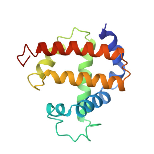
1.70 A resolution structure of myoglobin from yellowfin tuna. An example of a myoglobin lacking the D helix.

Birnbaum, G.I.Evans, S.V.Przybylska, M.Rose, D.R.

(1994) Acta Crystallogr D Biol Crystallogr 50: 283-289

  • DOI: https://doi.org/10.1107/S0907444993014271
  • Primary Citation of Related Structures:  
    1MYT

  • PubMed Abstract: 

    The crystal structure of metmyoglobin from yellowfin tuna (Thunnus albacares) has been determined by molecular replacement methods and refined to a conventional R factor of 0.177 for all observed reflections in the range of 6.0-1.70 A resolution. Like other myoglobins for which a high-resolution structure is available, the polypeptide chain is organized into several helices that cooperate to form a hydrophobic pocket into which the heme prosthetic group is non-covalently bound; however, the D helix observed in other myoglobins is absent in myoglobin from yellowfin tuna and has been replaced with a random coil. As well, the A helix has a pronounced kink due to the presence of Pro16. The differences in structure between this and sperm whale myoglobin can be correlated with their reported dioxygen affinity and dissociation. The structure is in agreement with reported fluorescence data which show an increased Trp14.heme distance in yellowfin tuna compared to sperm whale myoglobin.


  • Organizational Affiliation

    Institute for Biological Sciences, National Research Council of Canada, Ottawa.


Macromolecules
Find similar proteins by:  (by identity cutoff)  |  3D Structure
Entity ID: 1
MoleculeChains Sequence LengthOrganismDetailsImage
MYOGLOBIN146Thunnus albacaresMutation(s): 0 
UniProt
Find proteins for P02205 (Thunnus albacares)
Explore P02205 
Go to UniProtKB:  P02205
Entity Groups  
Sequence Clusters30% Identity50% Identity70% Identity90% Identity95% Identity100% Identity
UniProt GroupP02205
Sequence Annotations
Expand
  • Reference Sequence
Small Molecules
Ligands 1 Unique
IDChains Name / Formula / InChI Key2D Diagram3D Interactions
HEM
Query on HEM

Download Ideal Coordinates CCD File 
B [auth A]PROTOPORPHYRIN IX CONTAINING FE
C34 H32 Fe N4 O4
KABFMIBPWCXCRK-RGGAHWMASA-L
Experimental Data & Validation

Experimental Data

  • Method: X-RAY DIFFRACTION
  • Resolution: 1.74 Å
  • R-Value Observed: 0.177 
  • Space Group: P 21 21 21
Unit Cell:
Length ( Å )Angle ( ˚ )
a = 44.497α = 90
b = 72.318β = 90
c = 52.221γ = 90
Software Package:
Software NamePurpose
PROLSQrefinement

Structure Validation

View Full Validation Report


Ligand Structure Quality Assessment 


Entry History 

Deposition Data

Revision History  (Full details and data files)

  • Version 1.0: 1993-10-31
    Type: Initial release
  • Version 1.1: 2008-03-24
    Changes: Version format compliance
  • Version 1.2: 2011-07-13
    Changes: Version format compliance
  • Version 1.3: 2024-02-14
    Changes: Data collection, Database references, Derived calculations, Other