1LQW

Crystal Structure of S.aureus Peptide Deformylase


Experimental Data Snapshot

  • Method: X-RAY DIFFRACTION
  • Resolution: 1.87 Å
  • R-Value Free: 0.265 
  • R-Value Work: 0.231 
  • R-Value Observed: 0.231 

wwPDB Validation   3D Report Full Report


This is version 1.3 of the entry. See complete history


Literature

The crystal structures of four peptide deformylases bound to the antibiotic actinonin reveal two distinct types: a platform for the structure-based design of antibacterial agents.

Guilloteau, J.P.Mathieu, M.Giglione, C.Blanc, V.Dupuy, A.Chevrier, M.Gil, P.Famechon, A.Meinnel, T.Mikol, V.

(2002) J Mol Biol 320: 951-962

  • DOI: https://doi.org/10.1016/s0022-2836(02)00549-1
  • Primary Citation of Related Structures:  
    1LQW, 1LQY, 1LRU, 1LRY

  • PubMed Abstract: 

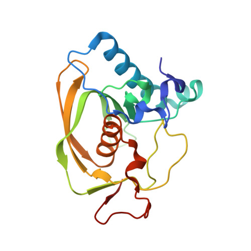
    Bacterial peptide deformylase (PDF) belongs to a sub-family of metalloproteases that catalyse the removal of the N-terminal formyl group from newly synthesised proteins. PDF is essential in prokaryotes and conserved throughout the eubacteria. It is therefore considered an attractive target for developing new antibacterial agents. Here, we report the crystal structures of four bacterial deformylases, free or bound to the naturally occurring antibiotic actinonin, including two from the major bacterial pathogens Pseudomonas aeruginosa and Staphylococcus aureus. The overall tertiary structure is essentially conserved but shows significant differences, namely at the C terminus, which are directly related to the deformylase type (i.e. I or II) they belong to. The geometry around the catalytic metal ion exhibits a high level of similarity within the different enzymes, as does the binding mode of actinonin to the various deformylases. However, some significant structural differences are found in the vicinity of the active site, highlighting the structural and molecular requirements for the design of a deformylase inhibitor active against a broad spectrum of bacterial strains.


  • Organizational Affiliation

    Drug Innovation & Approval, Aventis Pharma, 13 Quai Jules Guesde, BP.14, F-94403, Vitry-sur-Seine, France.


Macromolecules
Find similar proteins by:  (by identity cutoff)  |  3D Structure
Entity ID: 1
MoleculeChains Sequence LengthOrganismDetailsImage
PEPTIDE DEFORMYLASE PDF1
A, B
183Staphylococcus aureusMutation(s): 0 
EC: 3.5.1.88
UniProt
Find proteins for P68826 (Staphylococcus aureus)
Explore P68826 
Go to UniProtKB:  P68826
Entity Groups  
Sequence Clusters30% Identity50% Identity70% Identity90% Identity95% Identity100% Identity
UniProt GroupP68826
Sequence Annotations
Expand
  • Reference Sequence
Small Molecules
Ligands 1 Unique
IDChains Name / Formula / InChI Key2D Diagram3D Interactions
ZN
Query on ZN

Download Ideal Coordinates CCD File 
C [auth A],
D [auth B]
ZINC ION
Zn
PTFCDOFLOPIGGS-UHFFFAOYSA-N
Experimental Data & Validation

Experimental Data

  • Method: X-RAY DIFFRACTION
  • Resolution: 1.87 Å
  • R-Value Free: 0.265 
  • R-Value Work: 0.231 
  • R-Value Observed: 0.231 
  • Space Group: P 1 21 1
Unit Cell:
Length ( Å )Angle ( ˚ )
a = 48.21α = 90
b = 42.19β = 98.67
c = 101.67γ = 90
Software Package:
Software NamePurpose
AMoREphasing
X-PLORrefinement
MOSFLMdata reduction
CCP4data scaling

Structure Validation

View Full Validation Report


Entry History 

Deposition Data

  • Released Date: 2002-07-24 
  • Deposition Author(s): Mikol, V.

Revision History  (Full details and data files)

  • Version 1.0: 2002-07-24
    Type: Initial release
  • Version 1.1: 2008-04-28
    Changes: Version format compliance
  • Version 1.2: 2011-07-13
    Changes: Version format compliance
  • Version 1.3: 2024-02-14
    Changes: Data collection, Database references, Derived calculations