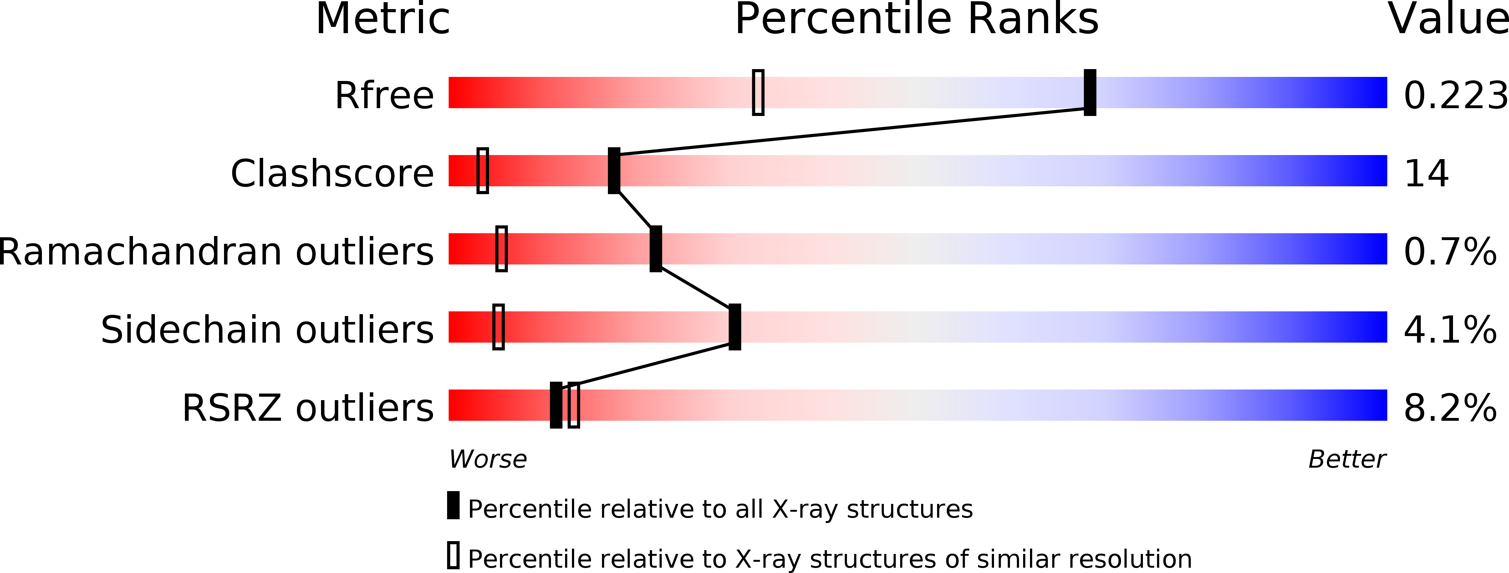
1IHR

Crystal structure of the dimeric C-terminal domain of TonB


Experimental Data Snapshot

  • Method: X-RAY DIFFRACTION
  • Resolution: 1.55 Å
  • R-Value Free: 0.230 
  • R-Value Work: 0.160 
  • R-Value Observed: 0.166 

wwPDB Validation   3D Report Full Report


This is version 1.3 of the entry. See complete history


Literature

Crystal structure of the dimeric C-terminal domain of TonB reveals a novel fold.

Chang, C.Mooser, A.Pluckthun, A.Wlodawer, A.

(2001) J Biol Chem 276: 27535-27540

  • DOI: https://doi.org/10.1074/jbc.M102778200
  • Primary Citation of Related Structures:  
    1IHR

  • PubMed Abstract: 

    The TonB-dependent complex of Gram-negative bacteria couples the inner membrane proton motive force to the active transport of iron.siderophore and vitamin B(12) across the outer membrane. The structural basis of that process has not been described so far in full detail. The crystal structure of the C-terminal domain of TonB from Escherichia coli has now been solved by multiwavelength anomalous diffraction and refined at 1.55-A resolution, providing the first evidence that this region of TonB (residues 164-239) dimerizes. Moreover, the structure shows a novel architecture that has no structural homologs among any known proteins. The dimer of the C-terminal domain of TonB is cylinder-shaped with a length of 65 A and a diameter of 25 A. Each monomer contains three beta strands and a single alpha helix. The two monomers are intertwined with each other, and all six beta-strands of the dimer make a large antiparallel beta-sheet. We propose a plausible model of binding of TonB to FhuA and FepA, two TonB-dependent outer-membrane receptors.


  • Organizational Affiliation

    Macromolecular Crystallography Laboratory, NCI, National Institutes of Health, Frederick, Maryland 21702, USA.


Macromolecules
Find similar proteins by:  (by identity cutoff)  |  3D Structure
Entity ID: 1
MoleculeChains Sequence LengthOrganismDetailsImage
TonB protein
A, B
76Escherichia coliMutation(s): 0 
Gene Names: JM83
UniProt
Find proteins for P02929 (Escherichia coli (strain K12))
Explore P02929 
Go to UniProtKB:  P02929
Entity Groups  
Sequence Clusters30% Identity50% Identity70% Identity90% Identity95% Identity100% Identity
UniProt GroupP02929
Sequence Annotations
Expand
  • Reference Sequence
Experimental Data & Validation

Experimental Data

  • Method: X-RAY DIFFRACTION
  • Resolution: 1.55 Å
  • R-Value Free: 0.230 
  • R-Value Work: 0.160 
  • R-Value Observed: 0.166 
  • Space Group: P 21 21 2
Unit Cell:
Length ( Å )Angle ( ˚ )
a = 63.77α = 90
b = 86.33β = 90
c = 26.55γ = 90
Software Package:
Software NamePurpose
SOLVEphasing
SHELXL-97refinement
DENZOdata reduction
SCALEPACKdata scaling

Structure Validation

View Full Validation Report


Entry History 

Deposition Data

Revision History  (Full details and data files)

  • Version 1.0: 2001-08-01
    Type: Initial release
  • Version 1.1: 2008-04-27
    Changes: Version format compliance
  • Version 1.2: 2011-07-13
    Changes: Version format compliance
  • Version 1.3: 2024-02-07
    Changes: Data collection, Database references, Derived calculations