1IDR

CRYSTAL STRUCTURE OF THE TRUNCATED-HEMOGLOBIN-N FROM MYCOBACTERIUM TUBERCULOSIS


Experimental Data Snapshot

  • Method: X-RAY DIFFRACTION
  • Resolution: 1.90 Å
  • R-Value Free: 0.250 
  • R-Value Work: 0.189 
  • R-Value Observed: 0.192 

wwPDB Validation   3D Report Full Report


Ligand Structure Quality Assessment 


This is version 1.3 of the entry. See complete history


Literature

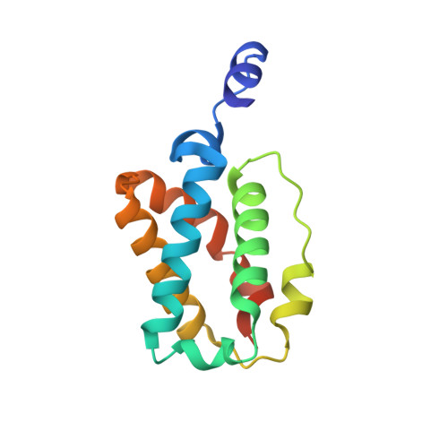
Mycobacterium tuberculosis hemoglobin N displays a protein tunnel suited for O2 diffusion to the heme.

Milani, M.Pesce, A.Ouellet, Y.Ascenzi, P.Guertin, M.Bolognesi, M.

(2001) EMBO J 20: 3902-3909

  • DOI: https://doi.org/10.1093/emboj/20.15.3902
  • Primary Citation of Related Structures:  
    1IDR

  • PubMed Abstract: 

    Macrophage-generated oxygen- and nitrogen-reactive species control the development of Mycobacterium tuberculosis infection in the host. Mycobacterium tuberculosis 'truncated hemoglobin' N (trHbN) has been related to nitric oxide (NO) detoxification, in response to macrophage nitrosative stress, during the bacterium latent infection stage. The three-dimensional structure of oxygenated trHbN, solved at 1.9 A resolution, displays the two-over-two alpha-helical sandwich fold recently characterized in two homologous truncated hemoglobins, featuring an extra N-terminal alpha-helix and homodimeric assembly. In the absence of a polar distal E7 residue, the O2 heme ligand is stabilized by two hydrogen bonds to TyrB10(33). Strikingly, ligand diffusion to the heme in trHbN may occur via an apolar tunnel/cavity system extending for approximately 28 A through the protein matrix, connecting the heme distal cavity to two distinct protein surface sites. This unique structural feature appears to be conserved in several homologous truncated hemoglobins. It is proposed that in trHbN, heme Fe/O2 stereochemistry and the protein matrix tunnel may promote O2/NO chemistry in vivo, as a M.tuberculosis defense mechanism against macrophage nitrosative stress.


  • Organizational Affiliation

    Department of Physics-INFM and Advanced Biotechnology Center-IST, University of Genova, Largo Rosanna Benzi 10. 16132 Genova, Italy.


Macromolecules
Find similar proteins by:  (by identity cutoff)  |  3D Structure
Entity ID: 1
MoleculeChains Sequence LengthOrganismDetailsImage
HEMOGLOBIN HBN
A, B
136Mycobacterium tuberculosisMutation(s): 0 
UniProt
Find proteins for P9WN25 (Mycobacterium tuberculosis (strain ATCC 25618 / H37Rv))
Explore P9WN25 
Go to UniProtKB:  P9WN25
Entity Groups  
Sequence Clusters30% Identity50% Identity70% Identity90% Identity95% Identity100% Identity
UniProt GroupP9WN25
Sequence Annotations
Expand
  • Reference Sequence
Experimental Data & Validation

Experimental Data

  • Method: X-RAY DIFFRACTION
  • Resolution: 1.90 Å
  • R-Value Free: 0.250 
  • R-Value Work: 0.189 
  • R-Value Observed: 0.192 
  • Space Group: P 21 21 21
Unit Cell:
Length ( Å )Angle ( ˚ )
a = 44.8α = 90
b = 62.248β = 90
c = 91.02γ = 90
Software Package:
Software NamePurpose
AMoREphasing
REFMACrefinement
DENZOdata reduction
SCALEPACKdata scaling

Structure Validation

View Full Validation Report


Ligand Structure Quality Assessment 


Entry History 

Deposition Data

Revision History  (Full details and data files)

  • Version 1.0: 2001-08-22
    Type: Initial release
  • Version 1.1: 2008-04-27
    Changes: Version format compliance
  • Version 1.2: 2011-07-13
    Changes: Version format compliance
  • Version 1.3: 2024-02-07
    Changes: Data collection, Database references, Derived calculations