1GG3

CRYSTAL STRUCTURE OF THE PROTEIN 4.1R MEMBRANE BINDING DOMAIN


Experimental Data Snapshot

  • Method: X-RAY DIFFRACTION
  • Resolution: 2.80 Å
  • R-Value Free: 0.276 
  • R-Value Work: 0.226 
  • R-Value Observed: 0.226 

wwPDB Validation   3D Report Full Report


This is version 1.3 of the entry. See complete history


Literature

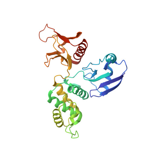
Protein 4.1R core domain structure and insights into regulation of cytoskeletal organization.

Han, B.G.Nunomura, W.Takakuwa, Y.Mohandas, N.Jap, B.K.

(2000) Nat Struct Biol 7: 871-875

  • DOI: https://doi.org/10.1038/82819
  • Primary Citation of Related Structures:  
    1GG3

  • PubMed Abstract: 

    The crystal structure of the core domain (N-terminal 30 kDa domain) of cytoskeletal protein 4.1R has been determined and shows a cloverleaf-like architecture. Each lobe of the cloverleaf contains a specific binding site for either band 3, glycophorin C/D or p55. At a central region of the molecule near where the three lobes are joined are two separate calmodulin (CaM) binding regions. One of these is composed primarily of an alpha-helix and is Ca 2+ insensitive; the other takes the form of an extended structure and its binding with CaM is dramatically enhanced by the presence of Ca 2+, resulting in the weakening of protein 4.1R binding to its target proteins. This novel architecture, in which the three lobes bind with three membrane associated proteins, and the location of calmodulin binding sites provide insight into how the protein 4.1R core domain interacts with membrane proteins and dynamically regulates cell shape in response to changes in intracellular Ca2+ levels.


  • Organizational Affiliation

    Life Sciences Division, Lawrence Berkeley National Laboratory, University of California, Berkeley, California 94720, USA.


Macromolecules
Find similar proteins by:  (by identity cutoff)  |  3D Structure
Entity ID: 1
MoleculeChains Sequence LengthOrganismDetailsImage
ERYTHROID MEMBRANE PROTEIN 4.1R
A, B, C
279Homo sapiensMutation(s): 0 
UniProt & NIH Common Fund Data Resources
Find proteins for P11171 (Homo sapiens)
Explore P11171 
Go to UniProtKB:  P11171
PHAROS:  P11171
GTEx:  ENSG00000159023 
Entity Groups  
Sequence Clusters30% Identity50% Identity70% Identity90% Identity95% Identity100% Identity
UniProt GroupP11171
Sequence Annotations
Expand
  • Reference Sequence
Experimental Data & Validation

Experimental Data

  • Method: X-RAY DIFFRACTION
  • Resolution: 2.80 Å
  • R-Value Free: 0.276 
  • R-Value Work: 0.226 
  • R-Value Observed: 0.226 
  • Space Group: C 1 2 1
Unit Cell:
Length ( Å )Angle ( ˚ )
a = 163.9α = 90
b = 106.5β = 95.5
c = 93.5γ = 90
Software Package:
Software NamePurpose
SOLVEphasing
CNSrefinement
DENZOdata reduction
SCALEPACKdata scaling

Structure Validation

View Full Validation Report


Entry History 

Deposition Data

  • Released Date: 2001-01-10 
  • Deposition Author(s): Han, B.G.

Revision History  (Full details and data files)

  • Version 1.0: 2001-01-10
    Type: Initial release
  • Version 1.1: 2008-04-26
    Changes: Version format compliance
  • Version 1.2: 2011-07-13
    Changes: Version format compliance
  • Version 1.3: 2023-12-27
    Changes: Data collection, Database references