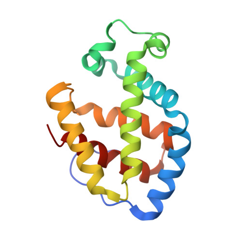
Crystalline ligand transitions in lamprey hemoglobin. Structural evidence for the regulation of oxygen affinity.
Heaslet, H.A., Royer Jr., W.E.(2001) J Biological Chem 276: 26230-26236
- PubMed: 11340069
- DOI: https://doi.org/10.1074/jbc.M101391200
- Primary Citation of Related Structures:
1F5O, 1F5P - PubMed Abstract:
The hemoglobins of the Sea Lamprey (Petromyzon marinus) exist in an equilibrium between low affinity oligomers, stabilized by proton binding, and higher affinity monomers, stabilized by oxygen binding. Recent crystallographic analysis revealed that dimerization is coupled with key changes at the ligand binding site with the distal histidine sterically restricting ligand binding in the deoxy dimer but with no significant structural rearrangements on the proximal side. These structural insights led to the hypothesis that oxygen affinity of lamprey hemoglobin is distally regulated. Here we present the 2.9-A crystal structure of deoxygenated lamprey hemoglobin in an orthorhombic crystal form along with the structure of these crystals exposed to carbon monoxide. The hexameric assemblage in this crystal form is very similar to those observed in the previous deoxy structure. Whereas the hydrogen bonding network and packing contacts formed in the dimeric interface of lamprey hemoglobin are largely unaffected by ligand binding, the binding of carbon monoxide induces the distal histidine to swing to positions that would preclude the formation of a stabilizing hydrogen bond with the bound ligand. These results suggest a dual role for the distal histidine and strongly support the hypothesis that ligand affinity in lamprey hemoglobin is distally regulated.
- Department of Biochemistry and Molecular Pharmacology, University of Massachusetts Medical School, Worcester, Massachusetts 01655, USA.
Organizational Affiliation: