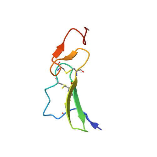
Three-dimensional nuclear magnetic resonance structures of mouse epidermal growth factor in acidic and physiological pH solutions.
Kohda, D., Inagaki, F.(1992) Biochemistry 31: 11928-11939
- PubMed: 1445923
- DOI: https://doi.org/10.1021/bi00162a036
- Primary Citation of Related Structures:
1EPG, 1EPH, 1EPI, 1EPJ - PubMed Abstract:
The three-dimensional structures of epidermal growth factors (EGF) previously reported were all in acidic solutions (pH 2.0-3.2), at which pHs EGF cannot bind to the receptor. Here we studied the structure of mouse EGF at pH 6.8, where EGF is physiologically active, and compared it with the structure at pH 2.0 by CD and NMR. From pH dependence of CD spectra and a comparison between the chemical shifts of the proton resonances at pH 6.8 and 2.0, the conformations at two pHs were found to be nearly identical except for the C-terminal tail region. The three-dimensional structures at pH 6.8 and 2.0 were determined independently by a combination of two-dimensional 1H NMR and stimulated annealing calculations using the program XPLOR. The calculations were based on 261 distance constraints at pH 6.8 and 355 distance and 24 torsion angle constraints at pH 2.0. The conformational difference of the C-terminal domain (residues 33-50) was detected between the two structures, which were supported by CD and the chemical shift comparison. The positions of the side chains of Leu47, Arg48, Trp49, and Trp50 are changed probably by the effect of the deprotonation of Asp46. Considering the fact that Leu47 is essential in EGF binding to the receptor, this conformational difference may be important in receptor recognition.
- Department of Molecular Physiology, Tokyo Metropolitan Institute of Medical Science, Japan.
Organizational Affiliation: