1DKH

CRYSTAL STRUCTURE OF THE HEMOPHORE HASA, PH 6.5


Experimental Data Snapshot

  • Method: X-RAY DIFFRACTION
  • Resolution: 3.20 Å
  • R-Value Free: 0.288 
  • R-Value Work: 0.228 
  • R-Value Observed: 0.230 

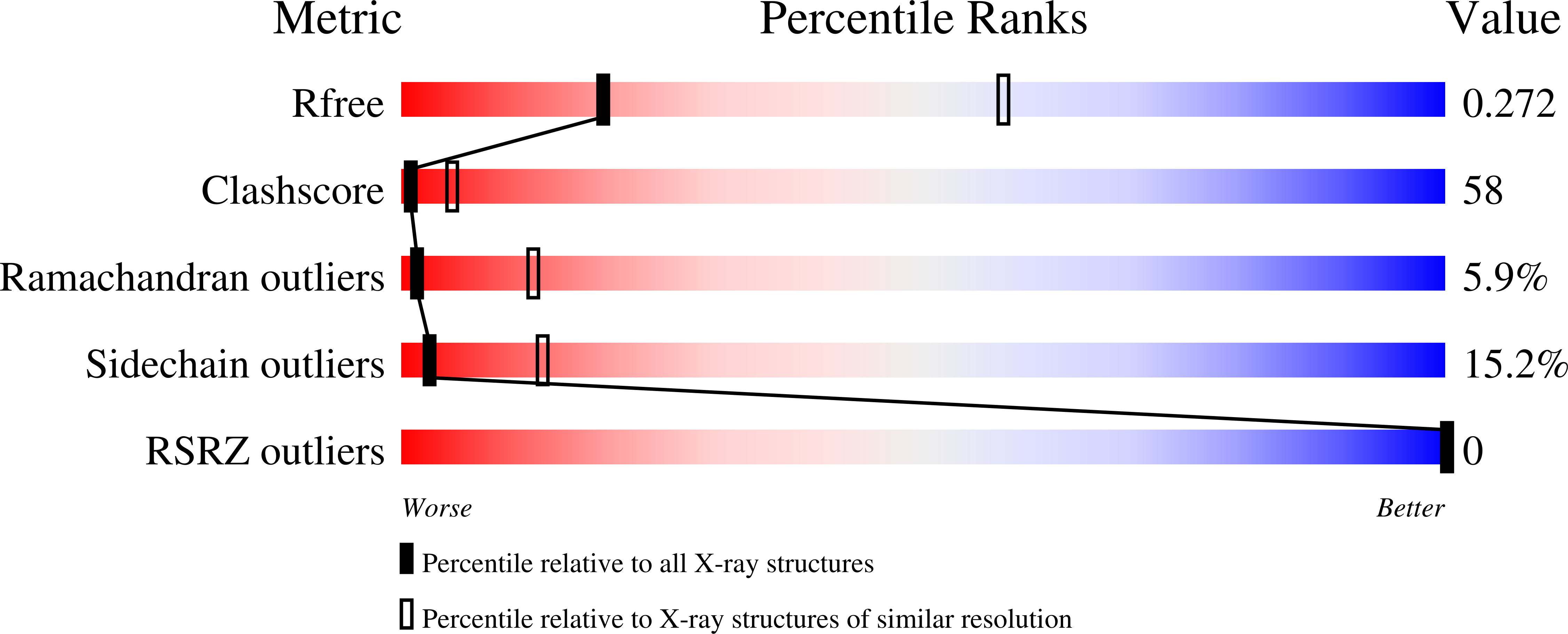
wwPDB Validation   3D Report Full Report


Ligand Structure Quality Assessment 


This is version 1.3 of the entry. See complete history


Literature

Functional aspects of the heme bound hemophore HasA by structural analysis of various crystal forms.

Arnoux, P.Haser, R.Izadi-Pruneyre, N.Lecroisey, A.Czjzek, M.

(2000) Proteins 41: 202-210

  • DOI: https://doi.org/10.1002/1097-0134(20001101)41:2<202::aid-prot50>3.0.co;2-8
  • Primary Citation of Related Structures:  
    1DK0, 1DKH

  • PubMed Abstract: 

    The protein HasA from the Gram negative bacteria Serratia marcescens is the first hemophore to be described at the molecular level. It participates to the shuttling of heme from hemoglobin to the outer membrane receptor HasR, which in turn releases it into the bacterium. HasR alone is also able to take up heme from hemoglobin but synergy with HasA increases the efficiency of the system by a factor of about 100. This iron acquisition system allows the bacteria to survive with hemoglobin as the sole iron source. Here we report the structures of a new crystal form of HasA diffracting up to 1.77A resolution as well as the refined structure of the trigonal crystal form diffracting to 3.2A resolution. The crystal structure of HasA at high resolution shows two possible orientations of the heme within the heme-binding pocket, which probably are functionally involved in the heme-iron acquisition process. The detailed analysis of the three known structures reveals the molecular basis regulating the relative affinity of the heme/hemophore complex.


  • Organizational Affiliation

    Laboratoire d'Architecture et Fonction des Macromolécules Biologiques, Institut de Biologie Structurale et Microbiologie, Marseille, France.


Macromolecules
Find similar proteins by:  (by identity cutoff)  |  3D Structure
Entity ID: 1
MoleculeChains Sequence LengthOrganismDetailsImage
HEME-BINDING PROTEIN A188Serratia marcescensMutation(s): 0 
UniProt
Find proteins for Q54450 (Serratia marcescens)
Explore Q54450 
Go to UniProtKB:  Q54450
Entity Groups  
Sequence Clusters30% Identity50% Identity70% Identity90% Identity95% Identity100% Identity
UniProt GroupQ54450
Sequence Annotations
Expand
  • Reference Sequence
Experimental Data & Validation

Experimental Data

  • Method: X-RAY DIFFRACTION
  • Resolution: 3.20 Å
  • R-Value Free: 0.288 
  • R-Value Work: 0.228 
  • R-Value Observed: 0.230 
  • Space Group: P 32 2 1
Unit Cell:
Length ( Å )Angle ( ˚ )
a = 111.65α = 90
b = 111.65β = 90
c = 52.79γ = 120
Software Package:
Software NamePurpose
SHARPphasing
CNSrefinement
DENZOdata reduction
CCP4data scaling

Structure Validation

View Full Validation Report


Ligand Structure Quality Assessment 


Entry History 

Deposition Data

Revision History  (Full details and data files)

  • Version 1.0: 2000-12-27
    Type: Initial release
  • Version 1.1: 2008-04-27
    Changes: Version format compliance
  • Version 1.2: 2011-07-13
    Changes: Version format compliance
  • Version 1.3: 2024-02-07
    Changes: Data collection, Database references, Derived calculations