Solution structure of a type I dockerin domain, a novel prokaryotic, extracellular calcium-binding domain.
Lytle, B.L., Volkman, B.F., Westler, W.M., Heckman, M.P., Wu, J.H.D.(2001) J Mol Biology 307: 745-753
- PubMed: 11273698
- DOI: https://doi.org/10.1006/jmbi.2001.4522
- Primary Citation of Related Structures:
1DAQ, 1DAV - PubMed Abstract:
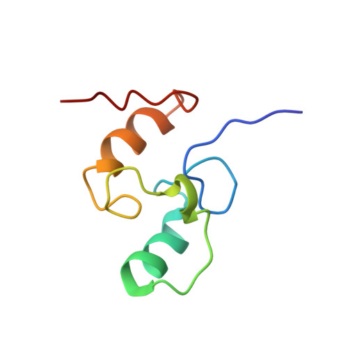
The type I dockerin domain is responsible for incorporating its associated glycosyl hydrolase into the bacterial cellulosome, a multienzyme cellulolytic complex, via its interaction with a receptor domain (cohesin domain) of the cellulosomal scaffolding subunit. The highly conserved dockerin domain is characterized by two Ca(2+)-binding sites with sequence similarity to the EF-hand motif. Here, we present the three-dimensional solution structure of the 69 residue dockerin domain of Clostridium thermocellum cellobiohydrolase CelS. Torsion angle dynamics calculations utilizing a total of 728 NOE-derived distance constraints and 79 torsion angle restraints yielded an ensemble of 20 structures with an average backbone r.m.s.d. for residues 5 to 29 and 32 to 66 of 0.54 A from the mean structure. The structure consists of two Ca(2+)-binding loop-helix motifs connected by a linker; the E helices entering each loop of the classical EF-hand motif are absent from the dockerin domain. Each dockerin Ca(2+)-binding subdomain is stabilized by a cluster of buried hydrophobic side-chains. Structural comparisons reveal that, in its non-complexed state, the dockerin fold displays a dramatic departure from that of Ca(2+)-bound EF-hand domains. A putative cohesin-binding surface, comprised of conserved hydrophobic and basic residues, is proposed, providing new insight into cellulosome assembly.
- Department of Chemical Engineering, University of Rochester, NY 14627-0166, USA.
Organizational Affiliation: