1D5E

The role of phenylalanine 8 in the stabilization of the S protein-S peptide interaction: Packing and cavities


Experimental Data Snapshot

  • Method: X-RAY DIFFRACTION
  • Resolution: 2.25 Å
  • R-Value Free: 0.231 
  • R-Value Work: 0.209 
  • R-Value Observed: 0.209 

wwPDB Validation   3D Report Full Report


This is version 2.0 of the entry. See complete history


Literature

Thermodynamic and structural studies of cavity formation in proteins suggest that loss of packing interactions rather than the hydrophobic effect dominates the observed energetics.

Ratnaparkhi, G.S.Varadarajan, R.

(2000) Biochemistry 39: 12365-12374

  • DOI: https://doi.org/10.1021/bi000775k
  • Primary Citation of Related Structures:  
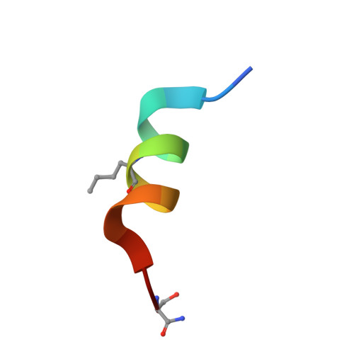
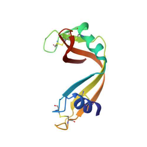
    1D5D, 1D5E, 1D5H

  • PubMed Abstract: 

    The hydrophobic effect is widely believed to be an important determinant of protein stability. However, it is difficult to obtain unambiguous experimental estimates of the contribution of the hydrophobic driving force to the overall free energy of folding. Thermodynamic and structural studies of large to small substitutions in proteins are the most direct method of measuring this contribution. We have substituted the buried residue Phe8 in RNase S with alanine, methionine, and norleucine. Binding thermodynamics and structures were characterized by titration calorimetry and crystallography, respectively. The crystal structures of the RNase S F8A, F8M, and F8Nle mutants indicate that the protein tolerates the changes without any main chain adjustments. The correlation of structural and thermodynamic parameters associated with large to small substitutions was analyzed for nine mutants of RNase S as well as 32 additional cavity-containing mutants of T4 lysozyme, human lysozyme, and barnase. Such substitutions were typically found to result in negligible changes in DeltaC(p)() and positive values of both DeltaDeltaH degrees and DeltaDeltaS of folding. Enthalpic effects were dominant, and the sign of DeltaDeltaS is the opposite of that expected from the hydrophobic effect. Values of DeltaDeltaG degrees and DeltaDeltaH degrees correlated better with changes in packing parameters such as residue depth or occluded surface than with the change in accessible surface area upon folding. These results suggest that the loss of packing interactions rather than the hydrophobic effect is a dominant contributor to the observed energetics for large to small substitutions. Hence, estimates of the magnitude of the hydrophobic driving force derived from earlier mutational studies are likely to be significantly in excess of the actual value.


  • Organizational Affiliation

    Molecular Biophysics Unit, Indian Institute of Science, Bangalore 560 012, India.


Macromolecules

Find similar proteins by:  Sequence   |   3D Structure  

Entity ID: 1
MoleculeChains Sequence LengthOrganismDetailsImage
S PEPTIDE15N/AMutation(s): 1 
UniProt
Find proteins for P61823 (Bos taurus)
Explore P61823 
Go to UniProtKB:  P61823
Entity Groups  
Sequence Clusters30% Identity50% Identity70% Identity90% Identity95% Identity100% Identity
UniProt GroupP61823
Sequence Annotations
Expand
  • Reference Sequence
Find similar proteins by:  (by identity cutoff)  |  3D Structure
Entity ID: 2
MoleculeChains Sequence LengthOrganismDetailsImage
RNASE S101Bos taurusMutation(s): 0 
UniProt
Find proteins for P61823 (Bos taurus)
Explore P61823 
Go to UniProtKB:  P61823
Entity Groups  
Sequence Clusters30% Identity50% Identity70% Identity90% Identity95% Identity100% Identity
UniProt GroupP61823
Sequence Annotations
Expand
  • Reference Sequence
Small Molecules
Ligands 1 Unique
IDChains Name / Formula / InChI Key2D Diagram3D Interactions
SO4
Query on SO4

Download Ideal Coordinates CCD File 
C [auth B]SULFATE ION
O4 S
QAOWNCQODCNURD-UHFFFAOYSA-L
Modified Residues  2 Unique
IDChains TypeFormula2D DiagramParent
NLE
Query on NLE
A
L-PEPTIDE LINKINGC6 H13 N O2LEU
SET
Query on SET
A
L-PEPTIDE LINKINGC3 H8 N2 O2SER
Experimental Data & Validation

Experimental Data

  • Method: X-RAY DIFFRACTION
  • Resolution: 2.25 Å
  • R-Value Free: 0.231 
  • R-Value Work: 0.209 
  • R-Value Observed: 0.209 
  • Space Group: P 31 2 1
Unit Cell:
Length ( Å )Angle ( ˚ )
a = 44.6α = 90
b = 44.6β = 90
c = 98.07γ = 120
Software Package:
Software NamePurpose
XDSdata scaling
AUTOMARdata reduction
X-PLORmodel building
X-PLORrefinement
XDSdata reduction
X-PLORphasing

Structure Validation

View Full Validation Report


Entry History 

Deposition Data

Revision History  (Full details and data files)

  • Version 1.0: 1999-10-20
    Type: Initial release
  • Version 1.1: 2008-04-27
    Changes: Version format compliance
  • Version 1.2: 2011-07-13
    Changes: Version format compliance
  • Version 1.3: 2017-10-04
    Changes: Refinement description
  • Version 1.4: 2018-03-14
    Changes: Database references
  • Version 1.5: 2018-04-04
    Changes: Data collection
  • Version 2.0: 2023-11-15
    Changes: Atomic model, Data collection, Database references, Derived calculations