1A7G

THE CRYSTAL STRUCTURE OF THE E2 DNA-BINDING DOMAIN FROM HUMAN PAPILLOMAVIRUS AT 2.4 ANGSTROMS


Experimental Data Snapshot

  • Method: X-RAY DIFFRACTION
  • Resolution: 2.40 Å
  • R-Value Free: 0.297 
  • R-Value Work: 0.211 
  • R-Value Observed: 0.211 

wwPDB Validation   3D Report Full Report


This is version 1.4 of the entry. See complete history


Literature

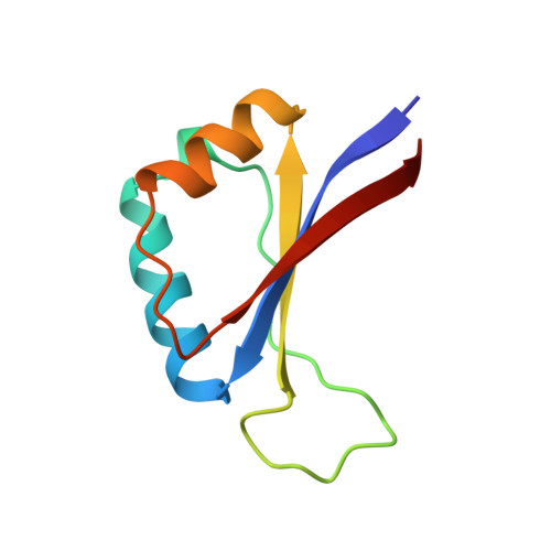
Structure of the E2 DNA-binding domain from human papillomavirus serotype 31 at 2.4 A.

Bussiere, D.E.Kong, X.Egan, D.A.Walter, K.Holzman, T.F.Lindh, F.Robins, T.Giranda, V.L.

(1998) Acta Crystallogr D Biol Crystallogr 54: 1367-1376

  • DOI: https://doi.org/10.1107/s0907444998005587
  • Primary Citation of Related Structures:  
    1A7G

  • PubMed Abstract: 

    The papillomaviruses are a family of small double-stranded DNA viruses which exclusively infect epithelial cells and stimulate the proliferation of those cells. A key protein within the papillomavirus life-cycle is known as the E2 (Early 2) protein and is responsible for regulating viral transcription from all viral promoters as well as for replication of the papillomavirus genome in tandem with another protein known as E1. The E2 protein itself consists of three functional domains: an N-terminal trans-activation domain, a proline-rich linker, and a C-terminal DNA-binding domain. The first crystal structure of the human papillomavirus, serotype 31 (HPV-31), E2 DNA-binding domain has been determined at 2.4 A resolution. The HPV DNA-binding domain monomer consists of two beta-alpha-beta repeats of approximately equal length and is arranged as to have an anti-parallel beta-sheet flanked by the two alpha-helices. The monomers form the functional in vivo dimer by association of the beta-sheets of each monomer so as to form an eight-stranded anti-parallel beta-barrel at the center of the dimer, with the alpha-helices lining the outside of the barrel. The overall structure of HVP-31 E2 DNA-binding domain is similar to both the bovine papillomavirus E2-binding domain and the Epstein-Barr nuclear antigen-1 DNA-binding domain.


  • Organizational Affiliation

    Division of Scientific Information, Analysis, and Management, Pharmaceutical Products Division, Abbott Laboratories, 100 Abbott Park Road, Abbott Park, IL 60064, USA.


Macromolecules
Find similar proteins by:  (by identity cutoff)  |  3D Structure
Entity ID: 1
MoleculeChains Sequence LengthOrganismDetailsImage
REGULATORY PROTEIN E2A [auth E]82human papillomavirus 31Mutation(s): 0 
Gene Names: E2
UniProt
Find proteins for P17383 (Human papillomavirus 31)
Explore P17383 
Go to UniProtKB:  P17383
Entity Groups  
Sequence Clusters30% Identity50% Identity70% Identity90% Identity95% Identity100% Identity
UniProt GroupP17383
Sequence Annotations
Expand
  • Reference Sequence
Small Molecules
Ligands 1 Unique
IDChains Name / Formula / InChI Key2D Diagram3D Interactions
SO4
Query on SO4

Download Ideal Coordinates CCD File 
B [auth E],
C [auth E]
SULFATE ION
O4 S
QAOWNCQODCNURD-UHFFFAOYSA-L
Experimental Data & Validation

Experimental Data

  • Method: X-RAY DIFFRACTION
  • Resolution: 2.40 Å
  • R-Value Free: 0.297 
  • R-Value Work: 0.211 
  • R-Value Observed: 0.211 
  • Space Group: P 61 2 2
Unit Cell:
Length ( Å )Angle ( ˚ )
a = 45.89α = 90
b = 45.89β = 90
c = 195.636γ = 120
Software Package:
Software NamePurpose
X-PLORmodel building
X-PLORrefinement
DENZOdata reduction
SCALEPACKdata scaling
X-PLORphasing

Structure Validation

View Full Validation Report


Entry History 

Deposition Data

Revision History  (Full details and data files)

  • Version 1.0: 1999-04-27
    Type: Initial release
  • Version 1.1: 2008-03-24
    Changes: Version format compliance
  • Version 1.2: 2011-07-13
    Changes: Derived calculations, Version format compliance
  • Version 1.3: 2024-02-07
    Changes: Data collection, Database references, Derived calculations, Other
  • Version 1.4: 2024-04-03
    Changes: Refinement description