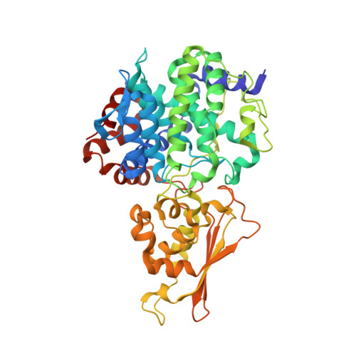
12VT | pdb_000012vt

Y318A Human Aconitate Decarboxylase 1 mutant, apo


Experimental Data Snapshot

  • Method: X-RAY DIFFRACTION
  • Resolution: 1.62 Å
  • R-Value Free: 
    0.202 (Depositor), 0.202 (DCC) 
  • R-Value Work: 
    0.160 (Depositor), 0.160 (DCC) 

Starting Model: experimental
View more details

wwPDB Validation 3D Report Full Report

Validation slider image for 12VT

This is version 1.1 of the entry. See complete history

Literature

Robust structural, kinetic and biophysical characterization of wild-type human ACOD1, selected mutants and their interaction with citraconate.

Runge, B.Oktay, H.Fucci, I.J.Merten, E.M.Tarasov, S.G.Fan, L.Monteiro, D.C.F.

(2026) J Struct Biol X 14: 100157-100157

  • DOI: https://doi.org/10.1016/j.yjsbx.2026.100157
  • Primary Citation Related Structures: 
    12UX, 12VD, 12VQ, 12VT, 12VW

  • PubMed Abstract: 

    Aconitate decarboxylase 1, an enzyme member of the MmgE-PrpD family of proteins, has gained significant attention in the last decade as a therapeutic target for cancer and inflammatory diseases. Its product, itaconate, is a multifunctional metabolite shown to drive several disease states. Though extensively studied in cellulo and in vivo , this protein is biochemically and mechanistically under characterized and although a family of inhibitors has been described, no ligand-bound structures have yet been determined. In this work we present a thorough structural investigation that yielded the first ligand-bound structure of this protein family, which required the generation of artifact-free apo crystals. We also developed a novel, low-consumption, robust kinetic assay and investigated active site and allosteric mutants to further elucidate structural and dynamic activity relationships of this protein.


  • Organizational Affiliation
    • Center for Structural Biology, Center for Cancer Research, National Cancer Institute, National Institutes of Health, Frederick, MD 21702, USA.

Macromolecule Content 

  • Total Structure Weight: 101.39 kDa 
  • Atom Count: 8,004 
  • Modeled Residue Count: 916 
  • Deposited Residue Count: 924 
  • Unique protein chains: 1

Macromolecules

Find similar proteins by:|  3D Structure
Entity ID: 1
MoleculeChains  Sequence LengthOrganismDetailsImage
Cis-aconitate decarboxylase
A, B
462Homo sapiensMutation(s): 1 
Gene Names: ACOD1IRG1
EC: 4.1.1.6
UniProt & NIH Common Fund Data Resources
Find proteins for A6NK06 (Homo sapiens)
Explore A6NK06 
Go to UniProtKB:  A6NK06
PHAROS:  A6NK06
GTEx:  ENSG00000102794 
Entity Groups
Sequence Clusters30% Identity50% Identity70% Identity90% Identity95% Identity100% Identity
UniProt GroupA6NK06
Sequence Annotations
Expand
Reference Sequence

Experimental Data & Validation

Experimental Data

  • Method: X-RAY DIFFRACTION
  • Resolution: 1.62 Å
  • R-Value Free:  0.202 (Depositor), 0.202 (DCC) 
  • R-Value Work:  0.160 (Depositor), 0.160 (DCC) 
Space Group: P 21 21 2
Unit Cell:
Length ( Å )Angle ( ˚ )
a = 101.889α = 90
b = 110.165β = 90
c = 76.11γ = 90
Software Package:
Software NamePurpose
REFMACrefinement
autoPROCdata reduction
STARANISOdata scaling
MOLREPphasing

Structure Validation

View Full Validation Report


Entry History 

& Funding Information

Deposition Data


Funding OrganizationLocationGrant Number
National Institutes of Health/National Cancer Institute (NIH/NCI)United States--

Revision History  (Full details and data files)

  • Version 1.0: 2026-08-19
    Type: Initial release
  • Version 1.1: 2026-09-02
    Changes: Database references