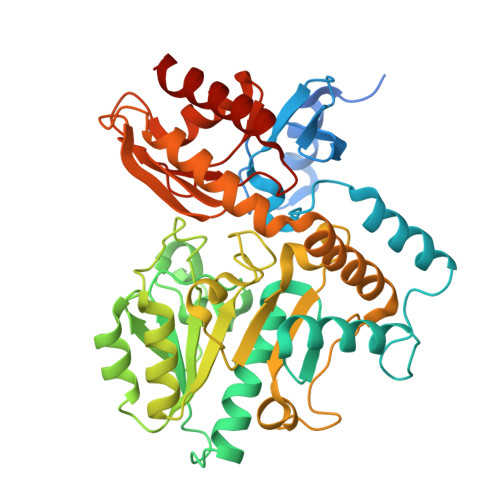
12AX | pdb_000012ax

Human Ornithine Aminotransferase soaked with its inhibitor, (3S,4R)-3-amino-4-ethynylcyclopent-1-ene-1-carboxylic acid


Experimental Data Snapshot

  • Method: X-RAY DIFFRACTION
  • Resolution: 1.58 Å
  • R-Value Free: 
    0.195 (Depositor), 0.194 (DCC) 
  • R-Value Work: 
    0.163 (Depositor), 0.163 (DCC) 
  • R-Value Observed: 
    0.164 (Depositor) 

Starting Model: experimental
View more details

wwPDB Validation 3D Report Full Report

Validation slider image for 12AX

This is version 1.0 of the entry. See complete history

Literature

Mechanism-Based Inactivation of Human Ornithine Aminotransferase by Ethynyl- and Nitrile-Substituted Cyclopentene Analogues of gamma-Aminobutyric Acids.

Wang, F.Corrigan, M.C.Le, N.H.V.Duan, D.Smith, C.O.Moran, G.R.Kelleher, N.L.Liu, D.Silverman, R.B.

(2026) J Am Chem Soc 148: 30301-30313

  • DOI: https://doi.org/10.1021/jacs.6c08456
  • Primary Citation Related Structures: 
    12AL, 12AV, 12AX, 12AY, 12AZ

  • PubMed Abstract: 

    Human ornithine aminotransferase ( h OAT), a pyridoxal 5'-phosphate (PLP)-dependent enzyme, plays a central role in glutamine, proline, and polyamine metabolism and is increasingly recognized as a metabolic vulnerability in multiple cancers. Previously, we established a second deprotonation strategy to achieve efficient mechanism-based inactivation of h OAT over closely related aminotransferases. Building on this concept, we report the rational design, synthesis, and mechanistic investigation of cyclopentene-based γ-aminobutyric acid analogues bearing alkyne or nitrile warheads as potent h OAT inactivators. These compounds undergo enzyme-catalyzed γ-deprotonation to form ketimine intermediates, priming for a subsequent tautomerization event that leads to irreversible inhibition. Inhibitory activity evaluation revealed pronounced stereochemical effects on binding affinity and partition ratio, with one nitrile analogue ( 4b ) exhibiting an exceptional inactivation efficiency ( k inact / K I = 111.8 mM -1 ·min -1 ) and ∼400-fold selectivity for h OAT over γ-aminobutyric acid aminotransferase. Intact protein mass spectrometry and X-ray crystallography demonstrated that alkyne-containing analogues form covalent adducts with h OAT, whereas nitrile-containing analogues generate noncovalent but tight-binding species. Kinetic isotope effect studies identified γ-deprotonation as the rate-determining step, and a complementary small-molecule mass and computational study elucidated the inactivation and turnover pathways. Collectively, these results expand the mechanistic repertoire of PLP-dependent enzyme inactivation and provide a generalizable framework for designing highly selective mechanism-based inactivators.


  • Organizational Affiliation
    • Department of Chemistry, Northwestern University, Evanston, Illinois 60208, United States.

Macromolecule Content 

  • Total Structure Weight: 147.85 kDa 
  • Atom Count: 10,748 
  • Modeled Residue Count: 1,211 
  • Deposited Residue Count: 1,317 
  • Unique protein chains: 1

Macromolecules

Find similar proteins by:|  3D Structure
Entity ID: 1
MoleculeChains  Sequence LengthOrganismDetailsImage
Ornithine aminotransferase, mitochondrial
A, B, C
439Homo sapiensMutation(s): 0 
Gene Names: OAT
EC: 2.6.1.13
UniProt & NIH Common Fund Data Resources
Find proteins for P04181 (Homo sapiens)
Explore P04181 
Go to UniProtKB:  P04181
PHAROS:  P04181
GTEx:  ENSG00000065154 
Entity Groups
Sequence Clusters30% Identity50% Identity70% Identity90% Identity95% Identity100% Identity
UniProt GroupP04181
Sequence Annotations
Expand
Reference Sequence

Small Molecules

Ligands 2 Unique
IDChains Name / Formula / InChI Key2D Diagram3D Interactions
A1DBA(
Subject of Investigation/LOI)

Query on A1DBA


Download:Ideal Coordinates CCD File
D [auth A],
I [auth B],
N [auth C]
4-ethenyl-3-[[2-methyl-3-oxidanyl-5-[[oxidanyl-bis(oxidanylidene)-$l^{6}-phosphanyl]oxymethyl]pyridin-4-yl]methylamino]cyclopenta-1,3-diene-1-carboxylic acid
C16 H19 N2 O7 P
VGFZQNFWUVIJGY-UHFFFAOYSA-N
GOL

Query on GOL


Download:Ideal Coordinates CCD File
E [auth A]
F [auth A]
G [auth A]
H [auth A]
J [auth B]
E [auth A],
F [auth A],
G [auth A],
H [auth A],
J [auth B],
K [auth B],
L [auth B],
M [auth B],
O [auth C],
P [auth C]
GLYCEROL
C3 H8 O3
PEDCQBHIVMGVHV-UHFFFAOYSA-N

Experimental Data & Validation

Experimental Data

  • Method: X-RAY DIFFRACTION
  • Resolution: 1.58 Å
  • R-Value Free:  0.195 (Depositor), 0.194 (DCC) 
  • R-Value Work:  0.163 (Depositor), 0.163 (DCC) 
  • R-Value Observed: 0.164 (Depositor) 
Space Group: P 32 2 1
Unit Cell:
Length ( Å )Angle ( ˚ )
a = 115.837α = 90
b = 115.837β = 90
c = 186.331γ = 120
Software Package:
Software NamePurpose
PHENIXrefinement
autoPROCdata reduction
autoPROCdata scaling
PHENIXphasing

Structure Validation

View Full Validation Report


Entry History 

& Funding Information

Deposition Data


Funding OrganizationLocationGrant Number
National Institutes of Health/National Cancer Institute (NIH/NCI)United States--

Revision History  (Full details and data files)

  • Version 1.0: 2026-09-16
    Type: Initial release