10ED | pdb_000010ed

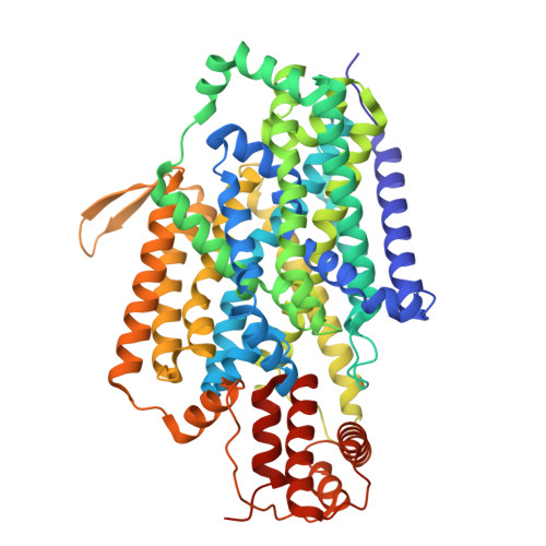
CbrXA SLC5-STAC domains


Experimental Data Snapshot

  • Method: ELECTRON MICROSCOPY
  • Resolution: 1.95 Å
  • Aggregation State: PARTICLE 
  • Reconstruction Method: SINGLE PARTICLE 

Starting Models: in silico
View more details

wwPDB Validation 3D Report Full Report

Validation slider image for 10ED

This is version 1.1 of the entry. See complete history

Literature

Structure and conformational dynamics of the Pseudomonas CbrA transceptor.

Orlando, M.A.Shah, T.Faber, M.W.Bose, S.Orlando, B.J.

(2026) Protein Sci 35: e70775-e70775

  • DOI: https://doi.org/10.1002/pro.70775
  • Primary Citation Related Structures: 
    10ED

  • PubMed Abstract: 

    The CbrA protein is a central regulator of carbon metabolism, biofilm formation, and virulence in Pseudomonas species, but the molecular mechanisms by which CbrA links nutrient sensing to downstream signaling has remained unclear. CbrA is a rare "transceptor" that combines membrane transporter and histidine kinase domains into a single functional polypeptide. The structural basis for histidine recognition and membrane transport, as well as signaling through intracellular histidine kinase domains has remained elusive. Here we determined a cryo-EM structure of CbrA which provides key molecular details of the SLC5-STAC domains in this unusual system. Unexpectedly, the small peptide CbrX encoded upstream of CbrA formed a stable complex with the SLC5 transporter domain, but was not essential for growth of Pseudomonas putida on histidine as a sole carbon source. The cryo-EM structure reveals how histidine binds within the transporter, and molecular dynamics simulations provide insight into proton gradient driven conformational changes that enable histidine transport. These findings define the molecular architecture of key CbrA functional domains, and lay the foundation for developing a comprehensive understanding of coupling between membrane transport and downstream signaling pathways that guide essential physiological traits in Pseudomonas.


  • Organizational Affiliation
    • Department of Biochemistry and Molecular Biology, Michigan State University, East Lansing, Michigan, USA.

Macromolecule Content 

  • Total Structure Weight: 79.23 kDa 
  • Atom Count: 5,448 
  • Modeled Residue Count: 647 
  • Deposited Residue Count: 664 
  • Unique protein chains: 2

Macromolecules

Find similar proteins by:|  3D Structure
Entity ID: 1
MoleculeChains  Sequence LengthOrganismDetailsImage
MFS transporterA [auth X]58Pseudomonas putida KT2440Mutation(s): 0 
Gene Names: PP_5704
UniProt
Find proteins for A0A140FWQ6 (Pseudomonas putida (strain ATCC 47054 / DSM 6125 / CFBP 8728 / NCIMB 11950 / KT2440))
Explore A0A140FWQ6 
Go to UniProtKB:  A0A140FWQ6
Entity Groups
Sequence Clusters30% Identity50% Identity70% Identity90% Identity95% Identity100% Identity
UniProt GroupA0A140FWQ6
Sequence Annotations
Expand
Reference Sequence
Find similar proteins by:|  3D Structure
Entity ID: 2
MoleculeChains  Sequence LengthOrganismDetailsImage
histidine kinaseB [auth A]606Pseudomonas putida KT2440Mutation(s): 0 
Gene Names: PP_4695
EC: 2.7.13.3
UniProt
Find proteins for Q88DX3 (Pseudomonas putida (strain ATCC 47054 / DSM 6125 / CFBP 8728 / NCIMB 11950 / KT2440))
Explore Q88DX3 
Go to UniProtKB:  Q88DX3
Entity Groups
Sequence Clusters30% Identity50% Identity70% Identity90% Identity95% Identity100% Identity
UniProt GroupQ88DX3
Sequence Annotations
Expand
Reference Sequence

Small Molecules

Ligands 3 Unique
IDChains Name / Formula / InChI Key2D Diagram3D Interactions
LMN

Query on LMN


Download:Ideal Coordinates CCD File
E [auth A],
F [auth A]
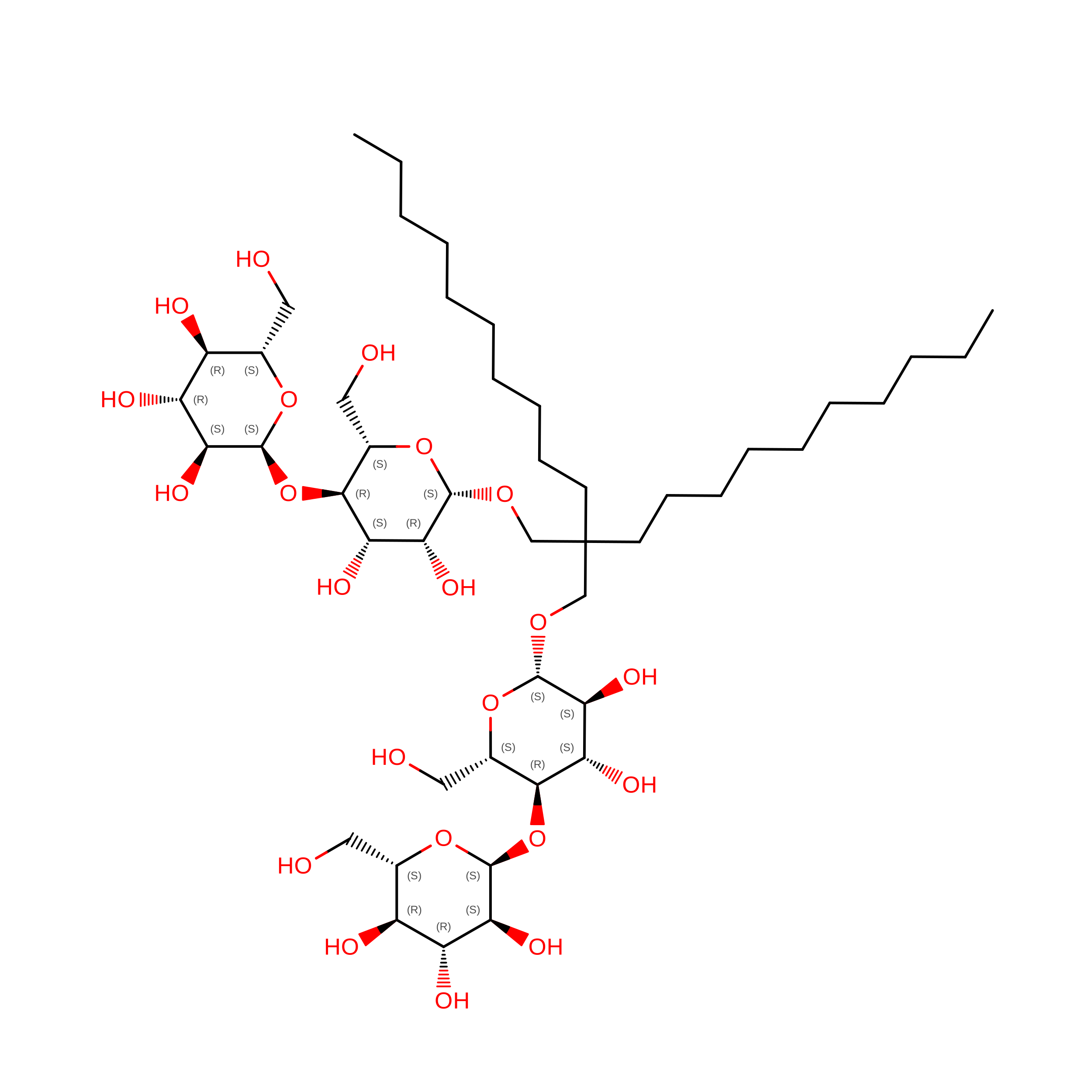
Lauryl Maltose Neopentyl Glycol
C47 H88 O22
MADJBYLAYPCCOO-XYPZXBMFSA-N
R16

Query on R16


Download:Ideal Coordinates CCD File
C [auth X]
G [auth A]
H [auth A]
I [auth A]
J [auth A]
C [auth X],
G [auth A],
H [auth A],
I [auth A],
J [auth A],
K [auth A],
L [auth A],
M [auth A],
N [auth A],
O [auth A],
P [auth A],
Q [auth A],
R [auth A],
S [auth A],
T [auth A]
HEXADECANE
C16 H34
DCAYPVUWAIABOU-UHFFFAOYSA-N
HIS
(Subject of Investigation/LOI)

Query on HIS


Download:Ideal Coordinates CCD File
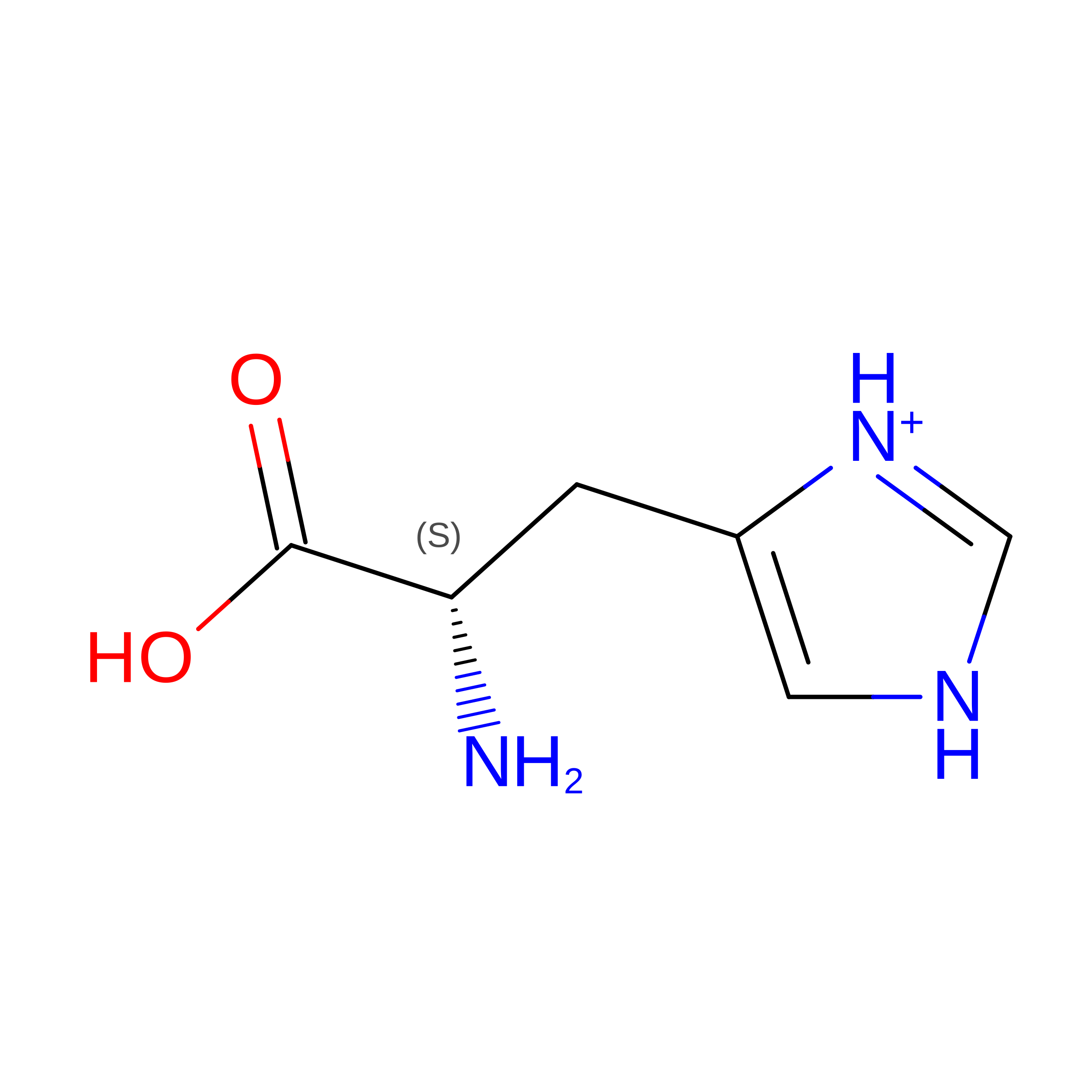
D [auth A]HISTIDINE
C6 H10 N3 O2
HNDVDQJCIGZPNO-YFKPBYRVSA-O

Experimental Data & Validation

Experimental Data

  • Method: ELECTRON MICROSCOPY
  • Resolution: 1.95 Å
  • Aggregation State: PARTICLE 
  • Reconstruction Method: SINGLE PARTICLE 
EM Software:
TaskSoftware PackageVersion
RECONSTRUCTIONcryoSPARC4.7.1
MODEL REFINEMENTPHENIX1.21.1_5286

Structure Validation

View Full Validation Report


Entry History 

& Funding Information

Deposition Data


Funding OrganizationLocationGrant Number
National Institutes of Health/National Institute of General Medical Sciences (NIH/NIGMS)United StatesR35 GM146721

Revision History  (Full details and data files)

  • Version 1.0: 2026-08-26
    Type: Initial release
  • Version 1.1: 2026-09-02
    Changes: Data collection, Database references