8FKB

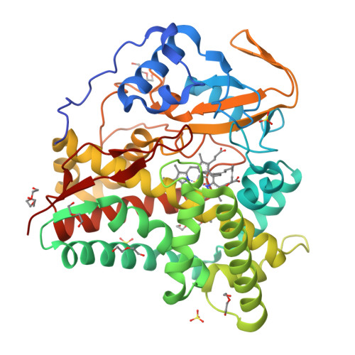
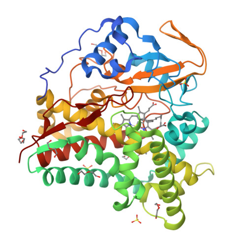
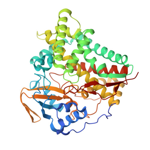
X-ray crystal structure of CYP124A1 from Mycobacterium Marinum bound to Farnesol


Experimental Data Snapshot

  • Method: X-RAY DIFFRACTION
  • Resolution: 1.42 Å
  • R-Value Free: 0.200 
  • R-Value Work: 0.183 
  • R-Value Observed: 0.183 

Starting Model: other
View more details

wwPDB Validation   3D Report Full Report


Ligand Structure Quality Assessment 


This is version 1.1 of the entry. See complete history


Literature

The catalytic activity and structure of the lipid metabolizing CYP124 cytochrome P450 enzyme from Mycobacterium marinum.

Ghith, A.Bruning, J.B.Bell, S.G.

(2023) Arch Biochem Biophys 737: 109554-109554

  • DOI: https://doi.org/10.1016/j.abb.2023.109554
  • Primary Citation of Related Structures:  
    8FJO, 8FKB, 8FLO

  • PubMed Abstract: 

    The CYP124 family of cytochrome P450 enzymes, as exemplified by CYP124A1 from Mycobacterium tuberculosis, is involved in the metabolism of methyl branched lipids and cholesterol derivatives. The equivalent enzyme from Mycobacterium marinum was investigated to compare the degree of functional conservation between members of this CYP family from closely related bacteria. We compared substrate binding of each CYP124 enzyme using UV-vis spectroscopy and the catalytic oxidation of methyl branched lipids, terpenes and cholesterol derivatives was investigated. The CYP124 enzyme from M. tuberculosis displayed a larger shift to the ferric high-spin state on binding cholesterol derivatives compared to the equivalent enzyme from M. marinum. The biggest difference was observed with cholesteryl sulfate which induced distinct UV-vis spectra in each CYP124 enzyme. The selectivity for oxidation at the ω-carbon of a branched chain was maintained for all substrates, except cholesteryl sulfate which was not oxidized by either enzyme. The CYP124A1 enzyme from M. marinum, in combination with farnesol and farnesyl acetate, was structurally characterized by X-ray crystallography. These ligand-bound structures of the CYP124 enzyme revealed that the polar component of the substrates bound in a different manner to that of phytanic acid in the structure of CYP124A1 from M. tuberculosis. However, closer to the heme the structures were similar providing an explanation for the high selectivity of the enzyme for terminal methyl C-H bond oxidation. The work here demonstrates that there were differences in the biochemistry of the CYP124 enzymes from these closely related bacteria.


  • Organizational Affiliation

    Department of Chemistry, University of Adelaide, SA, 5005, Australia.


Macromolecules
Find similar proteins by:  (by identity cutoff)  |  3D Structure
Entity ID: 1
MoleculeChains Sequence LengthOrganismDetailsImage
Cytochrome P450 124A1433Mycobacterium marinum ATCC BAA-535Mutation(s): 0 
Gene Names: cyp124A1
UniProt
Find proteins for B2HHT9 (Mycobacterium marinum (strain ATCC BAA-535 / M))
Explore B2HHT9 
Go to UniProtKB:  B2HHT9
Entity Groups  
Sequence Clusters30% Identity50% Identity70% Identity90% Identity95% Identity100% Identity
UniProt GroupB2HHT9
Sequence Annotations
Expand
  • Reference Sequence
Small Molecules
Ligands 5 Unique
IDChains Name / Formula / InChI Key2D Diagram3D Interactions
HEM
Query on HEM

Download Ideal Coordinates CCD File 
G [auth A]PROTOPORPHYRIN IX CONTAINING FE
C34 H32 Fe N4 O4
KABFMIBPWCXCRK-RGGAHWMASA-L
FOF (Subject of Investigation/LOI)
Query on FOF

Download Ideal Coordinates CCD File 
F [auth A](2E,6E)-3,7,11-trimethyldodeca-2,6,10-trien-1-ol
C15 H26 O
CRDAMVZIKSXKFV-YFVJMOTDSA-N
PGE
Query on PGE

Download Ideal Coordinates CCD File 
C [auth A],
H [auth A]
TRIETHYLENE GLYCOL
C6 H14 O4
ZIBGPFATKBEMQZ-UHFFFAOYSA-N
PEG
Query on PEG

Download Ideal Coordinates CCD File 
B [auth A],
D [auth A],
E [auth A],
I [auth A]
DI(HYDROXYETHYL)ETHER
C4 H10 O3
MTHSVFCYNBDYFN-UHFFFAOYSA-N
SO4
Query on SO4

Download Ideal Coordinates CCD File 
J [auth A],
K [auth A],
L [auth A]
SULFATE ION
O4 S
QAOWNCQODCNURD-UHFFFAOYSA-L
Experimental Data & Validation

Experimental Data

  • Method: X-RAY DIFFRACTION
  • Resolution: 1.42 Å
  • R-Value Free: 0.200 
  • R-Value Work: 0.183 
  • R-Value Observed: 0.183 
  • Space Group: C 1 2 1
Unit Cell:
Length ( Å )Angle ( ˚ )
a = 98.456α = 90
b = 72.425β = 109.56
c = 65.505γ = 90
Software Package:
Software NamePurpose
Aimlessdata scaling
PHENIXrefinement
XDSdata reduction
PHASERphasing

Structure Validation

View Full Validation Report


Ligand Structure Quality Assessment 


Entry History & Funding Information

Deposition Data


Funding OrganizationLocationGrant Number
Australian Research Council (ARC)AustraliaDP210103970

Revision History  (Full details and data files)

  • Version 1.0: 2023-03-29
    Type: Initial release
  • Version 1.1: 2024-05-22
    Changes: Data collection