7X4O

Crystal structure of Vps17p PX from S. cerevisiae (Space)


Experimental Data Snapshot

  • Method: X-RAY DIFFRACTION
  • Resolution: 2.02 Å
  • R-Value Free: 0.252 
  • R-Value Work: 0.200 
  • R-Value Observed: 0.202 

wwPDB Validation   3D Report Full Report


Ligand Structure Quality Assessment 


This is version 1.1 of the entry. See complete history


Literature

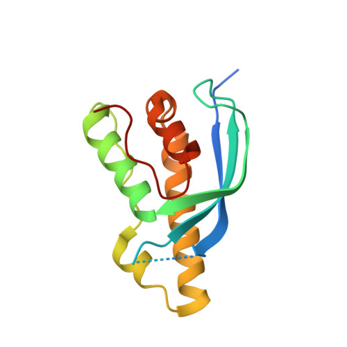
Crystal structure of the PX domain of Vps17p from Saccharomyces cerevisiae.

Obita, T.Inaka, K.Kohda, D.Maita, N.

(2022) Acta Crystallogr F Struct Biol Commun 78: 210-216

  • DOI: https://doi.org/10.1107/S2053230X22004472
  • Primary Citation of Related Structures:  
    7X4O

  • PubMed Abstract: 

    The structure determination of the PX (phox homology) domain of the Saccharomyces cerevisiae Vps17p protein presented a challenging case for molecular replacement because it has noncrystallographic symmetry close to a crystallographic axis. The combination of diffraction-quality crystals grown under microgravity on the International Space Station and a highly accurate template structure predicted by AlphaFold2 provided the key to successful crystal structure determination. Although the structure of the Vps17p PX domain is seen in many PX domains, no basic residues are found around the canonical phosphatidylinositol phosphate (PtdIns-P) binding site, suggesting an inability to bind PtdIns-P molecules.


  • Organizational Affiliation

    Faculty of Pharmaceutical Sciences, University of Toyama, Toyama 930-0194, Japan.


Macromolecules
Find similar proteins by:  (by identity cutoff)  |  3D Structure
Entity ID: 1
MoleculeChains Sequence LengthOrganismDetailsImage
Vacuolar protein sorting-associated protein 17
A, B
139Saccharomyces cerevisiae S288CMutation(s): 0 
Gene Names: VPS17PEP21YOR132WO3314YOR3314W
UniProt
Find proteins for P32913 (Saccharomyces cerevisiae (strain ATCC 204508 / S288c))
Explore P32913 
Go to UniProtKB:  P32913
Entity Groups  
Sequence Clusters30% Identity50% Identity70% Identity90% Identity95% Identity100% Identity
UniProt GroupP32913
Sequence Annotations
Expand
  • Reference Sequence
Experimental Data & Validation

Experimental Data

  • Method: X-RAY DIFFRACTION
  • Resolution: 2.02 Å
  • R-Value Free: 0.252 
  • R-Value Work: 0.200 
  • R-Value Observed: 0.202 
  • Space Group: P 21 3
Unit Cell:
Length ( Å )Angle ( ˚ )
a = 100.28α = 90
b = 100.28β = 90
c = 100.28γ = 90
Software Package:
Software NamePurpose
PHENIXrefinement
iMOSFLMdata reduction
Aimlessdata scaling
MOLREPphasing

Structure Validation

View Full Validation Report


Ligand Structure Quality Assessment 


Entry History & Funding Information

Deposition Data


Funding OrganizationLocationGrant Number
Japan Society for the Promotion of Science (JSPS)Japan16370050

Revision History  (Full details and data files)

  • Version 1.0: 2022-05-18
    Type: Initial release
  • Version 1.1: 2024-04-03
    Changes: Data collection, Refinement description