Crystal structure of Acetyl-CoA carboxylase (AccB) from Streptomyces antibioticus and insights into the substrate-binding through in silico mutagenesis and biophysical investigations.
Ali, I., Khan, A., Fa, Z., Khan, T., Wei, D.Q., Zheng, J.(2022) Comput Biol Med 145: 105439-105439
- PubMed: 35344865
- DOI: https://doi.org/10.1016/j.compbiomed.2022.105439
- Primary Citation of Related Structures:
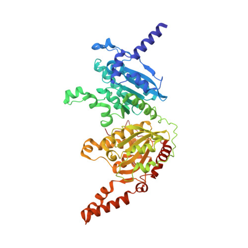
7W5U - PubMed Abstract:
Acetyl-CoA carboxylase (ACC) is crucial for polyketides biosynthesis and acts as an essential metabolic checkpoint. It is also an attractive drug target against obesity, cancer, microbial infections, and diabetes. However, the lack of knowledge, particularly sequence-structure function relationship to narrate ligand-enzyme binding, has hindered the progress of ACC-specific therapeutics and unnatural "natural" polyketides. Structural characterization of such enzymes will boost the opportunity to understand the substrate binding, designing new inhibitors and information regarding the molecular rules which control the substrate specificity of ACCs. To understand the substrate specificity, we determined the crystal structure of AccB (Carboxyl-transferase, CT) from Streptomyces antibioticus with a resolution of 2.3 Å and molecular modeling approaches were employed to unveil the molecular mechanism of acetyl-CoA recognition and processing. The CT domain of S. antibioticus shares a similar structural organization with the previous structures and the two steps reaction was confirmed by enzymatic assay. Furthermore, to reveal the key hotspots required for the substrate recognition and processing, in silico mutagenesis validated only three key residues (V223, Q346, and Q514) that help in the fixation of the substrate. Moreover, we also presented atomic level knowledge on the mechanism of the substrate binding, which unveiled the terminal loop (500-514) function as an opening and closing switch and pushes the substrate inside the cavity for stable binding. A significant decline in the hydrogen bonding half-life was observed upon the alanine substitution. Consequently, the presented structural data highlighted the potential key interacting residues for substrate recognition and will also help to re-design ACCs active site for proficient substrate specificity to produce diverse polyketides.
Organizational Affiliation:
State Key Laboratory of Microbial Metabolism, School of Life Sciences and Biotechnology, Shanghai Jiao Tong University, Shanghai, 200240, PR China.