7TZE

Structure of murine LAG3 domains 1-2

  • Classification: IMMUNOSUPPRESSANT
  • Organism(s): Mus musculus
  • Expression System: Trichoplusia ni
  • Mutation(s): No 

  • Deposited: 2022-02-15 Released: 2022-05-11 
  • Deposition Author(s): Ming, Q., Tran, T.H., Luca, V.C.
  • Funding Organization(s): National Institutes of Health/National Institute of General Medical Sciences (NIH/NIGMS), V Foundation for Cancer Research, Rita Allen Foundation

Experimental Data Snapshot

  • Method: X-RAY DIFFRACTION
  • Resolution: 2.12 Å
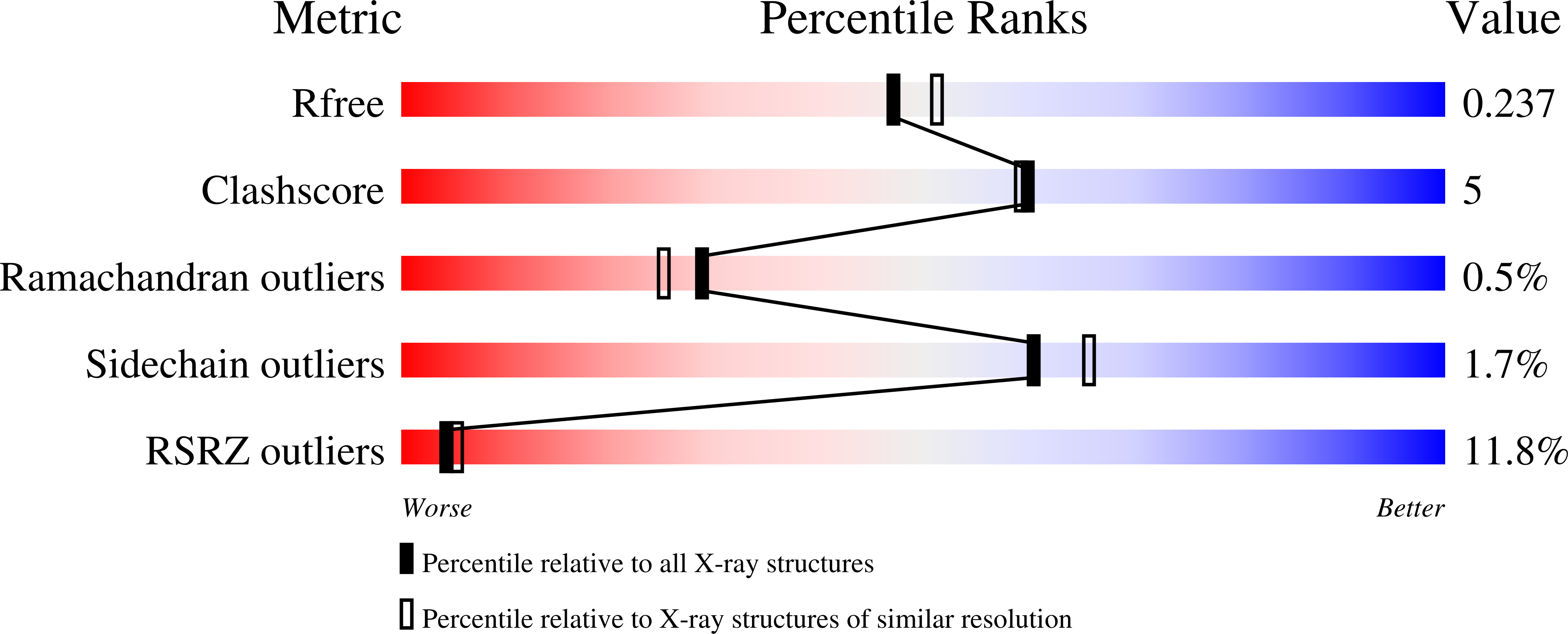
  • R-Value Free: 0.237 
  • R-Value Work: 0.212 
  • R-Value Observed: 0.213 

wwPDB Validation   3D Report Full Report


Ligand Structure Quality Assessment 


This is version 1.4 of the entry. See complete history


Literature

LAG3 ectodomain structure reveals functional interfaces for ligand and antibody recognition.

Ming, Q.Celias, D.P.Wu, C.Cole, A.R.Singh, S.Mason, C.Dong, S.Tran, T.H.Amarasinghe, G.K.Ruffell, B.Luca, V.C.

(2022) Nat Immunol 23: 1031-1041

  • DOI: https://doi.org/10.1038/s41590-022-01238-7
  • Primary Citation of Related Structures:  
    7TZ2, 7TZE, 7TZG, 7TZH

  • PubMed Abstract: 

    The immune checkpoint receptor lymphocyte activation gene 3 protein (LAG3) inhibits T cell function upon binding to major histocompatibility complex class II (MHC class II) or fibrinogen-like protein 1 (FGL1). Despite the emergence of LAG3 as a target for next-generation immunotherapies, we have little information describing the molecular structure of the LAG3 protein or how it engages cellular ligands. Here we determined the structures of human and murine LAG3 ectodomains, revealing a dimeric assembly mediated by Ig domain 2. Epitope mapping indicates that a potent LAG3 antagonist antibody blocks interactions with MHC class II and FGL1 by binding to a flexible 'loop 2' region in LAG3 domain 1. We also defined the LAG3-FGL1 interface by mapping mutations onto structures of LAG3 and FGL1 and established that FGL1 cross-linking induces the formation of higher-order LAG3 oligomers. These insights can guide LAG3-based drug development and implicate ligand-mediated LAG3 clustering as a mechanism for disrupting T cell activation.


  • Organizational Affiliation

    Department of Drug Discovery, Moffitt Cancer Center, Tampa, FL, USA.


Macromolecules
Find similar proteins by:  (by identity cutoff)  |  3D Structure
Entity ID: 1
MoleculeChains Sequence LengthOrganismDetailsImage
Lymphocyte activation gene 3 proteinA,
B [auth C]
232Mus musculusMutation(s): 0 
Gene Names: Lag3
UniProt
Find proteins for Q61790 (Mus musculus)
Explore Q61790 
Go to UniProtKB:  Q61790
Entity Groups  
Sequence Clusters30% Identity50% Identity70% Identity90% Identity95% Identity100% Identity
UniProt GroupQ61790
Sequence Annotations
Expand
  • Reference Sequence
Experimental Data & Validation

Experimental Data

  • Method: X-RAY DIFFRACTION
  • Resolution: 2.12 Å
  • R-Value Free: 0.237 
  • R-Value Work: 0.212 
  • R-Value Observed: 0.213 
  • Space Group: P 1 21 1
Unit Cell:
Length ( Å )Angle ( ˚ )
a = 35.729α = 90
b = 92.534β = 90.78
c = 81.771γ = 90
Software Package:
Software NamePurpose
XSCALEdata scaling
PHENIXrefinement
PDB_EXTRACTdata extraction
XDSdata reduction
PHASERphasing

Structure Validation

View Full Validation Report


Ligand Structure Quality Assessment 


Entry History & Funding Information

Deposition Data


Funding OrganizationLocationGrant Number
National Institutes of Health/National Institute of General Medical Sciences (NIH/NIGMS)United StatesR35GM133482
V Foundation for Cancer ResearchUnited States--
Rita Allen FoundationUnited States--

Revision History  (Full details and data files)

  • Version 1.0: 2022-05-11
    Type: Initial release
  • Version 1.1: 2022-05-18
    Changes: Structure summary
  • Version 1.2: 2022-07-13
    Changes: Database references
  • Version 1.3: 2022-07-27
    Changes: Database references
  • Version 1.4: 2023-10-18
    Changes: Data collection, Refinement description