Structure and function of an N-acetyltransferase from the human pathogen Acinetobacter baumannii isolate BAL_212.
Herkert, N.R., Thoden, J.B., Holden, H.M.(2022) Proteins 90: 1594-1605
- PubMed: 35277885
- DOI: https://doi.org/10.1002/prot.26334
- Primary Citation of Related Structures:
7TXP, 7TXQ, 7TXS - PubMed Abstract:
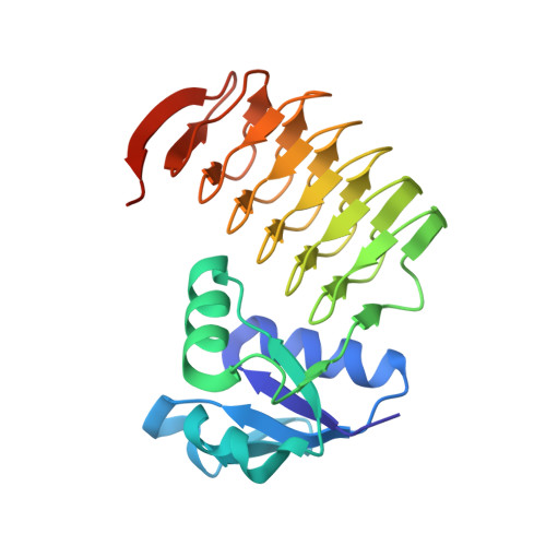
Acinetobacter baumannii is a Gram-negative bacterium commonly found in soil and water that can cause human infections of the blood, lungs, and urinary tract. Of particular concern is its prevalence in health-care settings where it can survive on surfaces and shared equipment for extended periods of time. The capsular polysaccharide surrounding the organism is known to be the major contributor to virulence. The structure of the K57 capsular polysaccharide produced by A. baumannii isolate BAL_212 from Vietnam was recently shown to contain the rare sugar 4-acetamido-4,6-dideoxy-d-glucose. Three enzymes are required for its biosynthesis, one of which is encoded by the gene H6W49_RS17300 and referred to as VioB, a putative N-acetyltransferase. Here, we describe a combined structural and functional analysis of VioB. Kinetic analyses show that the enzyme does, indeed, function on dTDP-4-amino-4,6-dideoxy-d-glucose with a catalytic efficiency of 3.9 x 10 4 M -1 s -1 (±6000), albeit at a reduced value compared to similar enzymes. Three high-resolution X-ray structures of various enzyme/ligand complexes were determined to resolutions of 1.65 Å or better. One of these models represents an intermediate analogue of the tetrahedral transition state. Differences between the VioB structure and those determined for the N-acetyltransferases from Campylobacter jejuni (PglD), Caulobacter crescentus (PerB), and Psychrobacter cryohalolentis (Pcryo_0637) are highlighted. Taken together, this investigation sheds new insight into the Type I sugar N-acetyltransferases.
Organizational Affiliation:
Department of Biochemistry, University of Wisconsin, Madison, Wisconsin, USA.