


Crystal structure of Shewanella benthica Group 1 truncated hemoglobin C51S C71S Y34F variant
Martinez, J.E., Liu, K., Siegler, M.A., Schlessman, J.L., Lecomte, J.T.J.To be published.
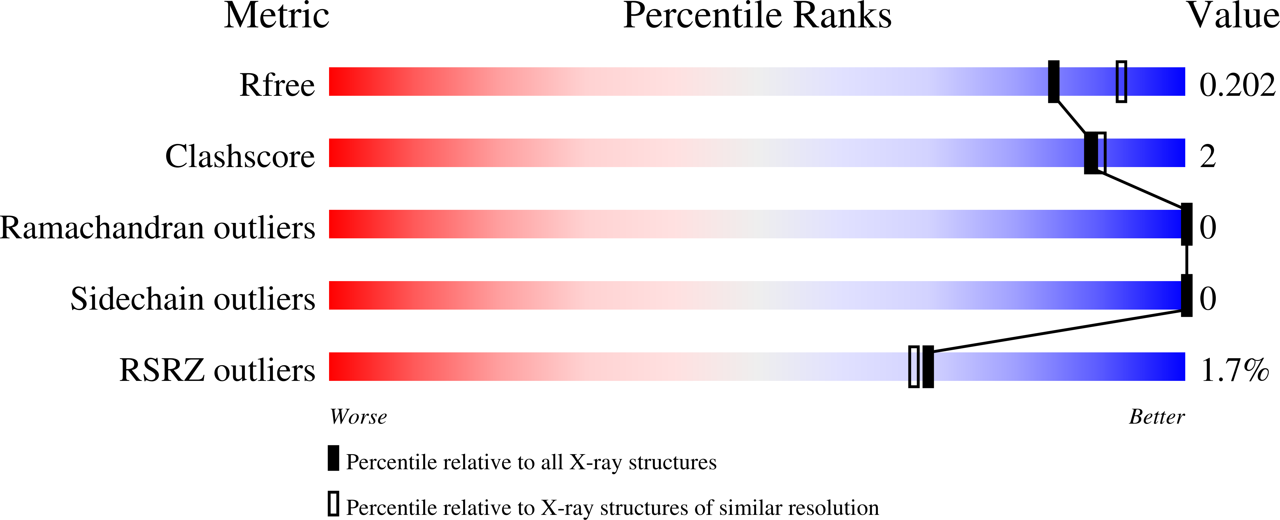
Experimental Data Snapshot
Entity ID: 1 | |||||
|---|---|---|---|---|---|
| Molecule | Chains | Sequence Length | Organism | Details | Image |
| Group 1 truncated hemoglobin | 116 | Shewanella benthica KT99 | Mutation(s): 3 Gene Names: KT99_16901 | 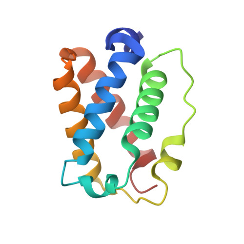 | |
UniProt | |||||
Find proteins for A9DF82 (Shewanella benthica KT99) Explore A9DF82 Go to UniProtKB: A9DF82 | |||||
Entity Groups | |||||
| Sequence Clusters | 30% Identity50% Identity70% Identity90% Identity95% Identity100% Identity | ||||
| UniProt Group | A9DF82 | ||||
Sequence AnnotationsExpand | |||||
| |||||
| Ligands 1 Unique | |||||
|---|---|---|---|---|---|
| ID | Chains | Name / Formula / InChI Key | 2D Diagram | 3D Interactions | |
| HEM (Subject of Investigation/LOI) Query on HEM | E [auth A], F [auth B], G [auth C], H [auth D] | PROTOPORPHYRIN IX CONTAINING FE C34 H32 Fe N4 O4 KABFMIBPWCXCRK-RGGAHWMASA-L |  | ||
| Length ( Å ) | Angle ( ˚ ) |
|---|---|
| a = 27.38 | α = 90 |
| b = 105.05 | β = 90 |
| c = 151.08 | γ = 90 |
| Software Name | Purpose |
|---|---|
| CrysalisPro | data reduction |
| CrysalisPro | data scaling |
| PHENIX | phasing |
| PHENIX | refinement |
| Coot | model building |
| Funding Organization | Location | Grant Number |
|---|---|---|
| National Science Foundation (NSF, United States) | United States | CHE-2003950 |
RCSB PDB (citation) is hosted by
RCSB PDB is a member of the

