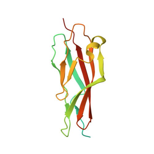
Functional and Structural Analysis of the Toxin-Binding Site of the Cadherin G-Protein-Coupled Receptor, BT-R 1 , for Cry1A Toxins of Bacillus thuringiensis .
Liu, L., Wilcox, X.E., Fisher, A.J., Boyd, S.D., Zhi, J., Winkler, D.D., Bulla Jr., L.A.(2022) Biochemistry 61: 752-766
- PubMed: 35438971
- DOI: https://doi.org/10.1021/acs.biochem.2c00089
- Primary Citation of Related Structures:
7TNI - PubMed Abstract:
The G-protein-coupled receptor BT-R 1 in the moth Manduca sexta represents a class of single-membrane-spanning α-helical proteins within the cadherin family that regulate intercellular adhesion and contribute to important signaling activities that control cellular homeostasis. The Cry1A toxins, Cry1Aa, Cry1Ab, and Cry1Ac, produced by Bacillus thuringiensis bind BT-R 1 very tightly ( K d = 1.1 nM) and trigger a Mg 2+ -dependent signaling pathway that involves the stimulation of G-protein α-subunit, which subsequently launches a coordinated signaling cascade, resulting in insect death. The three Cry1A toxins compete for the same binding site on BT-R 1 , and the pattern of inhibition of insecticidal activity against M. sexta is strikingly similar for all three toxins. The binding domain is localized in the 12th cadherin repeat (EC12: Asp1349 to Arg1460, 1349 DR 1460 ) in BT-R 1 and to various truncation fragments derived therefrom. Fine mapping of EC12 revealed that the smallest fragment capable of binding is a highly conserved 94-amino acid polypeptide bounded by Ile1363 and Ser1456 ( 1363 IS 1456 ), designated as the toxin-binding site (TBS). Logistical regression analysis revealed that binding of an EC12 truncation fragment containing the TBS is antagonistic to each of the Cry1A toxins and completely inhibits the insecticidal activity of all three. Elucidation of the EC12 motif of the TBS by X-ray crystallography at a 1.9 Å resolution combined with results of competitive binding analyses, live cell experiments, and whole insect bioassays substantiate the exclusive involvement of BT-R 1 in initiating insect cell death and demonstrate that the natural receptor BT-R 1 contains a single TBS.
Organizational Affiliation:
Department of Biological Sciences, The University of Texas at Dallas, Richardson, Texas 75083, United States.


