7TLF

Structure of the photoacclimated Light Harvesting Complex PE545 from Proteomonas sulcata


Experimental Data Snapshot

  • Method: X-RAY DIFFRACTION
  • Resolution: 2.80 Å
  • R-Value Free: 0.306 
  • R-Value Work: 0.245 
  • R-Value Observed: 0.248 

wwPDB Validation   3D Report Full Report


Ligand Structure Quality Assessment 


This is version 1.2 of the entry. See complete history


Literature

Controllable Phycobilin Modification: An Alternative Photoacclimation Response in Cryptophyte Algae.

Spangler, L.C.Yu, M.Jeffrey, P.D.Scholes, G.D.

(2022) ACS Cent Sci 8: 340-350

  • DOI: https://doi.org/10.1021/acscentsci.1c01209
  • Primary Citation of Related Structures:  
    7S96, 7S97, 7TJA, 7TLF

  • PubMed Abstract: 

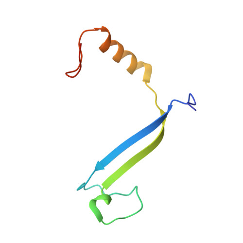
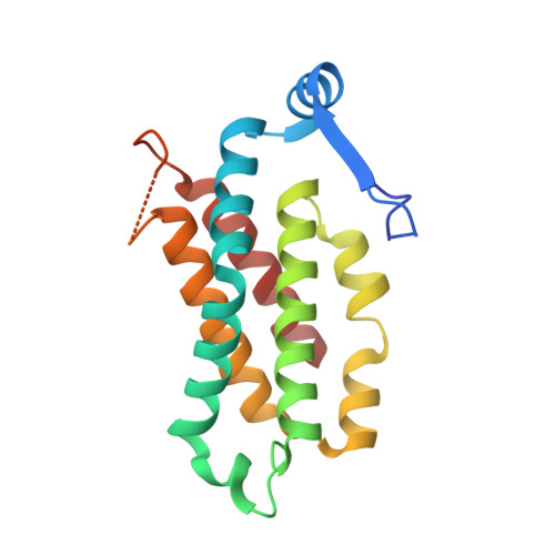
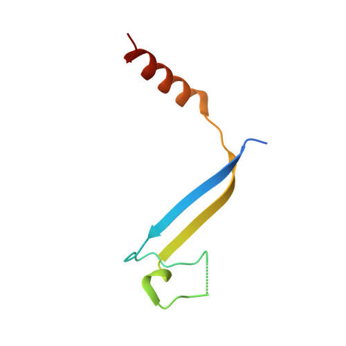
    Cryptophyte algae are well-known for their ability to survive under low light conditions using their auxiliary light harvesting antennas, phycobiliproteins. Mainly acting to absorb light where chlorophyll cannot (500-650 nm), phycobiliproteins also play an instrumental role in helping cryptophyte algae respond to changes in light intensity through the process of photoacclimation. Until recently, photoacclimation in cryptophyte algae was only observed as a change in the cellular concentration of phycobiliproteins; however, an additional photoacclimation response was recently discovered that causes shifts in the phycobiliprotein absorbance peaks following growth under red, blue, or green light. Here, we reproduce this newly identified photoacclimation response in two species of cryptophyte algae and elucidate the origin of the response on the protein level. We compare isolated native and photoacclimated phycobiliproteins for these two species using spectroscopy and mass spectrometry, and we report the X-ray structures of each phycobiliprotein and the corresponding photoacclimated complex. We find that neither the protein sequences nor the protein structures are modified by photoacclimation. We conclude that cryptophyte algae change one chromophore in the phycobiliprotein β subunits in response to changes in the spectral quality of light. Ultrafast pump-probe spectroscopy shows that the energy transfer is weakly affected by photoacclimation.


  • Organizational Affiliation

    Department of Chemistry, Princeton University, Princeton, New Jersey 08544, United States.


Macromolecules
Find similar proteins by:  (by identity cutoff)  |  3D Structure
Entity ID: 1
MoleculeChains Sequence LengthOrganismDetailsImage
Phycoerythrin alpha-subunit 1
A, E, I, M
76Proteomonas sulcataMutation(s): 0 
UniProt
Find proteins for A0A067YS87 (Proteomonas sulcata)
Explore A0A067YS87 
Go to UniProtKB:  A0A067YS87
Entity Groups  
Sequence Clusters30% Identity50% Identity70% Identity90% Identity95% Identity100% Identity
UniProt GroupA0A067YS87
Sequence Annotations
Expand
  • Reference Sequence
Find similar proteins by:  (by identity cutoff)  |  3D Structure
Entity ID: 2
MoleculeChains Sequence LengthOrganismDetailsImage
Phycoerythrin beta-subunit177Proteomonas sulcataMutation(s): 0 
Entity Groups  
Sequence Clusters30% Identity50% Identity70% Identity90% Identity95% Identity100% Identity
Sequence Annotations
Expand
  • Reference Sequence
Find similar proteins by:  (by identity cutoff)  |  3D Structure
Entity ID: 3
MoleculeChains Sequence LengthOrganismDetailsImage
Phycoerythrin alpha-subunit 2C,
F [auth G],
J [auth K],
N [auth O]
67Proteomonas sulcataMutation(s): 0 
UniProt
Find proteins for A0A067YRY3 (Proteomonas sulcata)
Explore A0A067YRY3 
Go to UniProtKB:  A0A067YRY3
Entity Groups  
Sequence Clusters30% Identity50% Identity70% Identity90% Identity95% Identity100% Identity
UniProt GroupA0A067YRY3
Sequence Annotations
Expand
  • Reference Sequence
Small Molecules
Ligands 2 Unique
IDChains Name / Formula / InChI Key2D Diagram3D Interactions
PEB (Subject of Investigation/LOI)
Query on PEB

Download Ideal Coordinates CCD File 
AA [auth F]
BA [auth F]
CA [auth F]
DA [auth H]
EA [auth H]
AA [auth F],
BA [auth F],
CA [auth F],
DA [auth H],
EA [auth H],
FA [auth H],
IA [auth J],
JA [auth J],
KA [auth J],
LA [auth L],
MA [auth L],
NA [auth L],
QA [auth N],
R [auth B],
RA [auth N],
S [auth B],
SA [auth N],
T [auth B],
TA [auth P],
UA [auth P],
V [auth D],
VA [auth P],
W [auth D],
X [auth D]
PHYCOERYTHROBILIN
C33 H40 N4 O6
NKCBCVIFPXGHAV-WAVSMFBNSA-N
DBV (Subject of Investigation/LOI)
Query on DBV

Download Ideal Coordinates CCD File 
GA [auth I]
HA [auth K]
OA [auth M]
PA [auth O]
Q [auth A]
GA [auth I],
HA [auth K],
OA [auth M],
PA [auth O],
Q [auth A],
U [auth C],
Y [auth E],
Z [auth G]
15,16-DIHYDROBILIVERDIN
C33 H36 N4 O6
ZQHDSLZHMAUUQK-ZTYGKHTCSA-N
Experimental Data & Validation

Experimental Data

  • Method: X-RAY DIFFRACTION
  • Resolution: 2.80 Å
  • R-Value Free: 0.306 
  • R-Value Work: 0.245 
  • R-Value Observed: 0.248 
  • Space Group: P 1 21 1
Unit Cell:
Length ( Å )Angle ( ˚ )
a = 88.249α = 90
b = 132.237β = 116.92
c = 93.413γ = 90
Software Package:
Software NamePurpose
PHENIXrefinement
XDSdata reduction
Aimlessdata scaling
PHASERphasing

Structure Validation

View Full Validation Report


Ligand Structure Quality Assessment 


Entry History & Funding Information

Deposition Data


Funding OrganizationLocationGrant Number
Canadian Institute for Advanced Research (CIFAR)Canada--

Revision History  (Full details and data files)

  • Version 1.0: 2022-02-09
    Type: Initial release
  • Version 1.1: 2022-04-20
    Changes: Database references
  • Version 1.2: 2023-10-18
    Changes: Data collection, Refinement description