Structure and Function of the beta-Asp-Arg Polymerase Cyanophycin Synthetase 2.
Sharon, I., Grogg, M., Hilvert, D., Schmeing, T.M.(2022) ACS Chem Biol 17: 670-679
- PubMed: 35179888
- DOI: https://doi.org/10.1021/acschembio.1c01007
- Primary Citation of Related Structures:
7TA5 - PubMed Abstract:
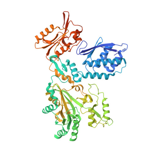
Cyanophycin is a biopolymer composed of long chains of β-Asp-Arg. It is widespread in nature, being synthesized by many clades of bacteria, which use it as a cellular reservoir of nitrogen, carbon, and energy. Two enzymes are known to produce cyanophycin: cyanophycin synthetase 1 (CphA1), which builds cyanophycin from the amino acids Asp and Arg by alternating between two separate reactions for backbone extension and side chain modification, and cyanophycin synthetase 2 (CphA2), which polymerizes β-Asp-Arg dipeptides. CphA2 is evolutionarily related to CphA1, but questions about CphA2's altered structure and function remain unresolved. Cyanophycin and related molecules have drawn interest as green biopolymers. Because it only has a single active site, CphA2 could be more useful than CphA1 for biotechnological applications seeking to produce modified cyanophycin. In this study, we report biochemical assays on nine cyanobacterial CphA2 enzymes and report the crystal structure of CphA2 from Gloeothece citriformis at 3.0 Å resolution. The structure reveals a homodimeric, three-domain architecture. One domain harbors the polymerization active site and the two other domains have structural roles. The structure and biochemical assays explain how CphA2 binds and polymerizes β-Asp-Arg and highlights differences in in vitro oligomerization and activity between CphA2 enzymes. Using the structure and distinct activity profile as a guide, we introduced a single point mutation that converted Gloeothece citriformis CphA2 from a primer-dependent enzyme into a primer-independent enzyme.
Organizational Affiliation:
Department of Biochemistry and Centre de recherche en biologie structurale, McGill University, Montréal H3G 0B1, Quebec, Canada.