7RKC

Computationally designed tunable C2 symmetric tandem repeat homodimer, D_3_633


Experimental Data Snapshot

  • Method: X-RAY DIFFRACTION
  • Resolution: 2.35 Å
  • R-Value Free: 0.312 
  • R-Value Work: 0.269 
  • R-Value Observed: 0.273 

wwPDB Validation   3D Report Full Report


This is version 1.2 of the entry. See complete history


Literature

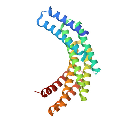
De novo design of protein homodimers containing tunable symmetric protein pockets.

Hicks, D.R.Kennedy, M.A.Thompson, K.A.DeWitt, M.Coventry, B.Kang, A.Bera, A.K.Brunette, T.J.Sankaran, B.Stoddard, B.Baker, D.

(2022) Proc Natl Acad Sci U S A 119: e2113400119-e2113400119

  • DOI: https://doi.org/10.1073/pnas.2113400119
  • Primary Citation of Related Structures:  
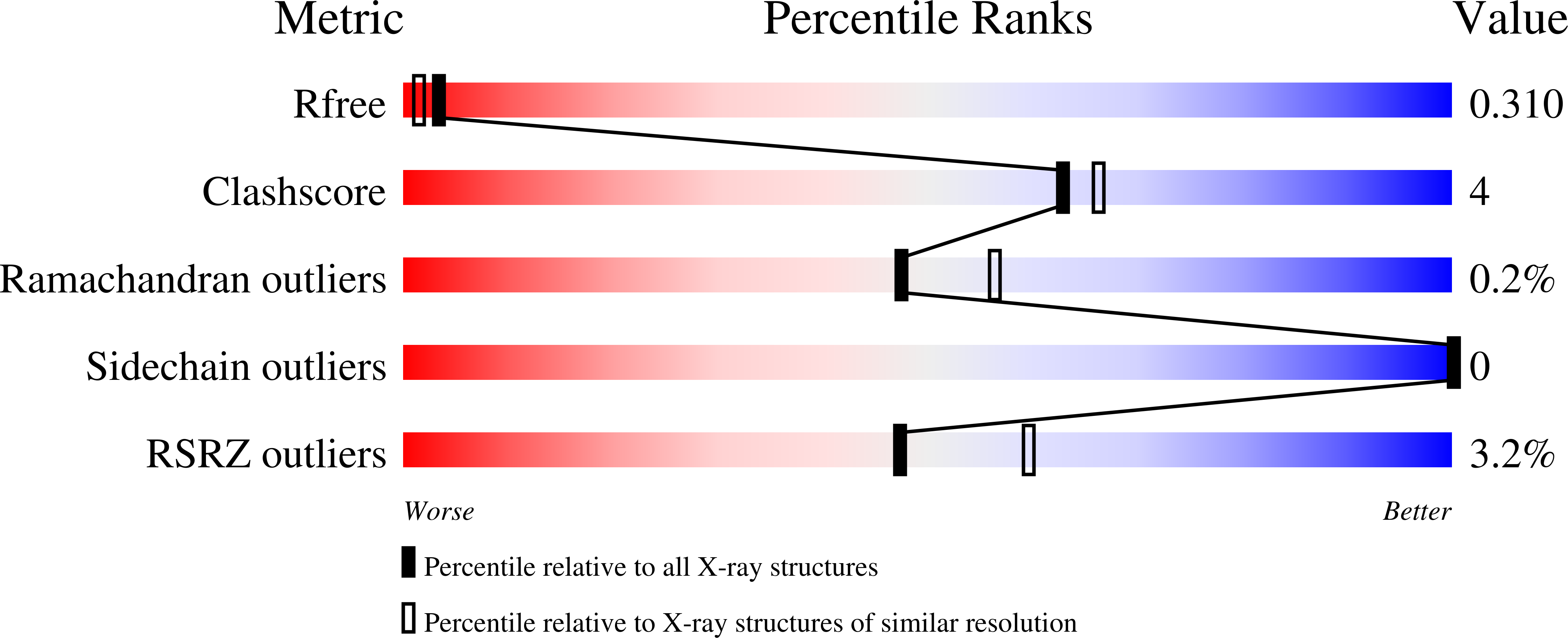
    7RKC, 7RMX, 7RMY

  • PubMed Abstract: 

    Function follows form in biology, and the binding of small molecules requires proteins with pockets that match the shape of the ligand. For design of binding to symmetric ligands, protein homo-oligomers with matching symmetry are advantageous as each protein subunit can make identical interactions with the ligand. Here, we describe a general approach to designing hyperstable C2 symmetric proteins with pockets of diverse size and shape. We first designed repeat proteins that sample a continuum of curvatures but have low helical rise, then docked these into C2 symmetric homodimers to generate an extensive range of C2 symmetric cavities. We used this approach to design thousands of C2 symmetric homodimers, and characterized 101 of them experimentally. Of these, the geometry of 31 were confirmed by small angle X-ray scattering and 2 were shown by crystallographic analyses to be in close agreement with the computational design models. These scaffolds provide a rich set of starting points for binding a wide range of C2 symmetric compounds.


  • Organizational Affiliation

    Department of Biochemistry, University of Washington, Seattle, WA 98195.


Macromolecules
Find similar proteins by:  (by identity cutoff)  |  3D Structure
Entity ID: 1
MoleculeChains Sequence LengthOrganismDetailsImage
D_3_633
A, B
233synthetic constructMutation(s): 0 
Entity Groups  
Sequence Clusters30% Identity50% Identity70% Identity90% Identity95% Identity100% Identity
Sequence Annotations
Expand
  • Reference Sequence
Experimental Data & Validation

Experimental Data

  • Method: X-RAY DIFFRACTION
  • Resolution: 2.35 Å
  • R-Value Free: 0.312 
  • R-Value Work: 0.269 
  • R-Value Observed: 0.273 
  • Space Group: P 1 21 1
Unit Cell:
Length ( Å )Angle ( ˚ )
a = 54.445α = 90
b = 54.682β = 95.47
c = 86.987γ = 90
Software Package:
Software NamePurpose
PHENIXrefinement
HKL-2000data scaling
PHASERphasing
PDB_EXTRACTdata extraction

Structure Validation

View Full Validation Report


Entry History & Funding Information

Deposition Data


Funding OrganizationLocationGrant Number
National Science Foundation (NSF, United States)United StatesDGE-1762114
National Institutes of Health/National Institute of General Medical Sciences (NIH/NIGMS)United StatesR01GM115545
National Institutes of Health/National Institute of General Medical Sciences (NIH/NIGMS)United StatesR01GM139752
National Institutes of Health/National Institute on Aging (NIH/NIA)United StatesR01AG063845
National Institutes of Health/National Institute on Aging (NIH/NIA)United StatesU19AG065156

Revision History  (Full details and data files)

  • Version 1.0: 2022-05-18
    Type: Initial release
  • Version 1.1: 2022-08-03
    Changes: Database references
  • Version 1.2: 2024-04-03
    Changes: Data collection, Refinement description