7RDS

Structure of human NTHL1


Experimental Data Snapshot

  • Method: X-RAY DIFFRACTION
  • Resolution: 2.50 Å
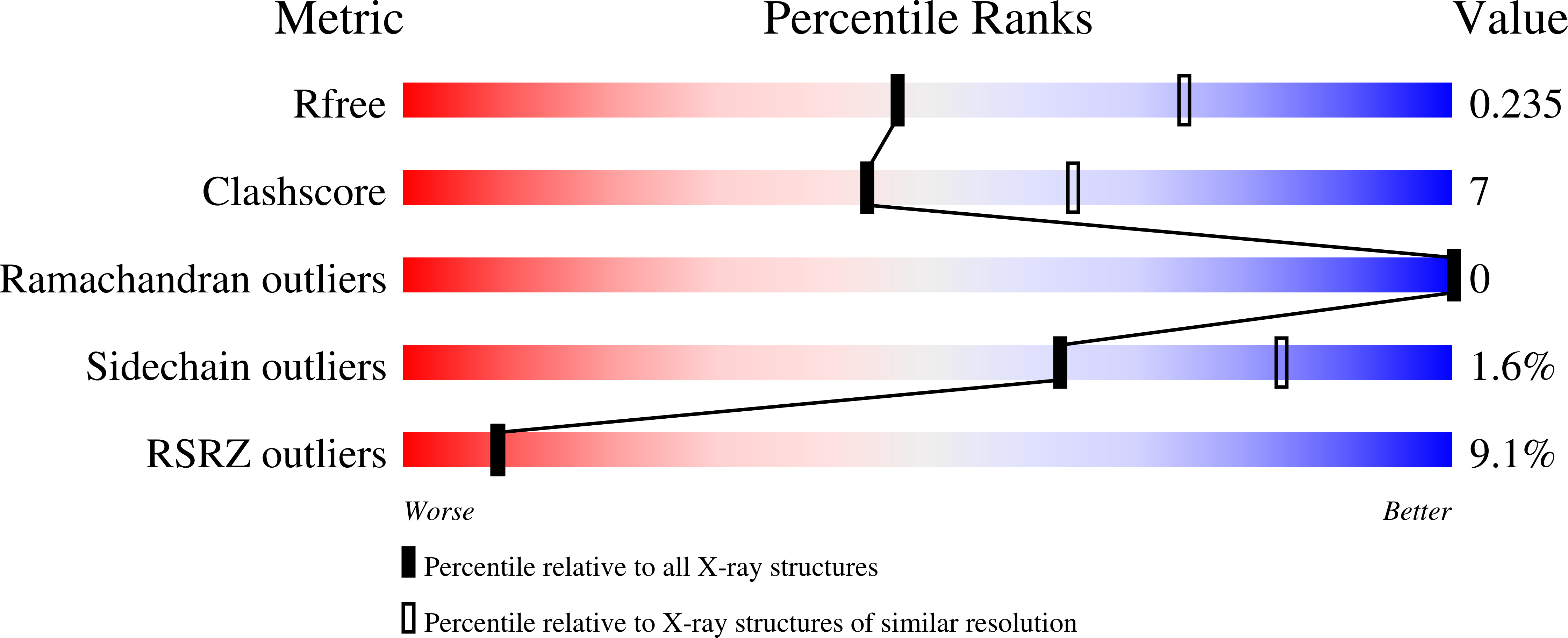
  • R-Value Free: 0.244 
  • R-Value Work: 0.200 
  • R-Value Observed: 0.202 

wwPDB Validation   3D Report Full Report


This is version 1.0 of the entry. See complete history


Literature

Caught in motion: human NTHL1 undergoes interdomain rearrangement necessary for catalysis.

Carroll, B.L.Zahn, K.E.Hanley, J.P.Wallace, S.S.Dragon, J.A.Doublie, S.

(2021) Nucleic Acids Res 49: 13165-13178

  • DOI: https://doi.org/10.1093/nar/gkab1162
  • Primary Citation of Related Structures:  
    7RDS, 7RDT

  • PubMed Abstract: 

    Base excision repair (BER) is the main pathway protecting cells from the continuous damage to DNA inflicted by reactive oxygen species. BER is initiated by DNA glycosylases, each of which repairs a particular class of base damage. NTHL1, a bifunctional DNA glycosylase, possesses both glycolytic and β-lytic activities with a preference for oxidized pyrimidine substrates. Defects in human NTHL1 drive a class of polyposis colorectal cancer. We report the first X-ray crystal structure of hNTHL1, revealing an open conformation not previously observed in the bacterial orthologs. In this conformation, the six-helical barrel domain comprising the helix-hairpin-helix (HhH) DNA binding motif is tipped away from the iron sulphur cluster-containing domain, requiring a conformational change to assemble a catalytic site upon DNA binding. We found that the flexibility of hNTHL1 and its ability to adopt an open configuration can be attributed to an interdomain linker. Swapping the human linker sequence for that of Escherichia coli yielded a protein chimera that crystallized in a closed conformation and had a reduced activity on lesion-containing DNA. This large scale interdomain rearrangement during catalysis is unprecedented for a HhH superfamily DNA glycosylase and provides important insight into the molecular mechanism of hNTHL1.


  • Organizational Affiliation

    Department of Microbiology and Molecular Genetics, University of Vermont, Burlington, VT 05405, USA.


Macromolecules
Find similar proteins by:  (by identity cutoff)  |  3D Structure
Entity ID: 1
MoleculeChains Sequence LengthOrganismDetailsImage
Isoform 3 of Endonuclease III-like protein 1257Homo sapiensMutation(s): 0 
Gene Names: NTHL1NTH1OCTS3
EC: 3.2.2 (PDB Primary Data), 4.2.99.18 (PDB Primary Data)
UniProt & NIH Common Fund Data Resources
Find proteins for P78549 (Homo sapiens)
Explore P78549 
Go to UniProtKB:  P78549
PHAROS:  P78549
GTEx:  ENSG00000065057 
Entity Groups  
Sequence Clusters30% Identity50% Identity70% Identity90% Identity95% Identity100% Identity
UniProt GroupP78549
Sequence Annotations
Expand
  • Reference Sequence
Small Molecules
Ligands 1 Unique
IDChains Name / Formula / InChI Key2D Diagram3D Interactions
SF4
Query on SF4

Download Ideal Coordinates CCD File 
B [auth A]IRON/SULFUR CLUSTER
Fe4 S4
LJBDFODJNLIPKO-UHFFFAOYSA-N
Experimental Data & Validation

Experimental Data

  • Method: X-RAY DIFFRACTION
  • Resolution: 2.50 Å
  • R-Value Free: 0.244 
  • R-Value Work: 0.200 
  • R-Value Observed: 0.202 
  • Space Group: P 63
Unit Cell:
Length ( Å )Angle ( ˚ )
a = 124.8α = 90
b = 124.8β = 90
c = 42.25γ = 120
Software Package:
Software NamePurpose
PHENIXrefinement
MOSFLMdata reduction
Aimlessdata scaling
PHENIXphasing

Structure Validation

View Full Validation Report


Entry History & Funding Information

Deposition Data


Funding OrganizationLocationGrant Number
National Institutes of Health/National Cancer Institute (NIH/NCI)United StatesP01-CA098993

Revision History  (Full details and data files)

  • Version 1.0: 2021-12-29
    Type: Initial release