7RCW

Crystal structure of C. difficile penicillin-binding protein 2 in complex with ampicillin


Experimental Data Snapshot

  • Method: X-RAY DIFFRACTION
  • Resolution: 3.00 Å
  • R-Value Free: 0.260 
  • R-Value Work: 0.197 
  • R-Value Observed: 0.200 

wwPDB Validation   3D Report Full Report


Ligand Structure Quality Assessment 


This is version 1.2 of the entry. See complete history


Literature

A unique class of Zn 2+ -binding serine-based PBPs underlies cephalosporin resistance and sporogenesis in Clostridioides difficile.

Sacco, M.D.Wang, S.Adapa, S.R.Zhang, X.Lewandowski, E.M.Gongora, M.V.Keramisanou, D.Atlas, Z.D.Townsend, J.A.Gatdula, J.R.Morgan, R.T.Hammond, L.R.Marty, M.T.Wang, J.Eswara, P.J.Gelis, I.Jiang, R.H.Y.Sun, X.Chen, Y.

(2022) Nat Commun 13: 4370-4370

  • DOI: https://doi.org/10.1038/s41467-022-32086-6
  • Primary Citation of Related Structures:  
    7RCW, 7RCX, 7RCY, 7RCZ, 7RD0

  • PubMed Abstract: 

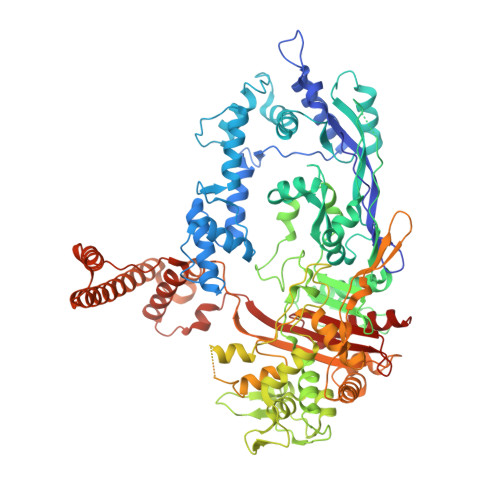
    Treatment with β-lactam antibiotics, particularly cephalosporins, is a major risk factor for Clostridioides difficile infection. These broad-spectrum antibiotics irreversibly inhibit penicillin-binding proteins (PBPs), which are serine-based enzymes that assemble the bacterial cell wall. However, C. difficile has four different PBPs (PBP1-3 and SpoVD) with various roles in growth and spore formation, and their specific links to β-lactam resistance in this pathogen are underexplored. Here, we show that PBP2 (known to be essential for vegetative growth) is the primary bactericidal target for β-lactams in C. difficile. PBP2 is insensitive to cephalosporin inhibition, and this appears to be the main basis for cephalosporin resistance in this organism. We determine crystal structures of C. difficile PBP2, alone and in complex with β-lactams, revealing unique features including ligand-induced conformational changes and an active site Zn 2+ -binding motif that influences β-lactam binding and protein stability. The Zn 2+ -binding motif is also present in C. difficile PBP3 and SpoVD (which are known to be essential for sporulation), as well as in other bacterial taxa including species living in extreme environments and the human gut. We speculate that this thiol-containing motif and its cognate Zn 2+ might function as a redox sensor to regulate cell wall synthesis for survival in adverse or anaerobic environments.


  • Organizational Affiliation

    Department of Molecular Medicine, Morsani College of Medicine, University of South Florida, Tampa, FL, 33612, USA.


Macromolecules
Find similar proteins by:  (by identity cutoff)  |  3D Structure
Entity ID: 1
MoleculeChains Sequence LengthOrganismDetailsImage
Penicillin-binding protein927Clostridioides difficile R20291Mutation(s): 0 
Gene Names: CDR20291_0985
UniProt
Find proteins for Q18B08 (Clostridioides difficile (strain 630))
Explore Q18B08 
Go to UniProtKB:  Q18B08
Entity Groups  
Sequence Clusters30% Identity50% Identity70% Identity90% Identity95% Identity100% Identity
UniProt GroupQ18B08
Sequence Annotations
Expand
  • Reference Sequence
Small Molecules
Ligands 5 Unique
IDChains Name / Formula / InChI Key2D Diagram3D Interactions
ZZ7 (Subject of Investigation/LOI)
Query on ZZ7

Download Ideal Coordinates CCD File 
E [auth A](2R,4S)-2-[(R)-{[(2R)-2-amino-2-phenylacetyl]amino}(carboxy)methyl]-5,5-dimethyl-1,3-thiazolidine-4-carboxylic acid
C16 H21 N3 O5 S
KDAWOPKDXRJNHV-MPPDQPJWSA-N
PEG
Query on PEG

Download Ideal Coordinates CCD File 
B [auth A]DI(HYDROXYETHYL)ETHER
C4 H10 O3
MTHSVFCYNBDYFN-UHFFFAOYSA-N
GOL
Query on GOL

Download Ideal Coordinates CCD File 
D [auth A]GLYCEROL
C3 H8 O3
PEDCQBHIVMGVHV-UHFFFAOYSA-N
ZN
Query on ZN

Download Ideal Coordinates CCD File 
F [auth A]ZINC ION
Zn
PTFCDOFLOPIGGS-UHFFFAOYSA-N
ACT
Query on ACT

Download Ideal Coordinates CCD File 
C [auth A]ACETATE ION
C2 H3 O2
QTBSBXVTEAMEQO-UHFFFAOYSA-M
Experimental Data & Validation

Experimental Data

  • Method: X-RAY DIFFRACTION
  • Resolution: 3.00 Å
  • R-Value Free: 0.260 
  • R-Value Work: 0.197 
  • R-Value Observed: 0.200 
  • Space Group: P 1 21 1
Unit Cell:
Length ( Å )Angle ( ˚ )
a = 62.91α = 90
b = 200.19β = 100.11
c = 70.68γ = 90
Software Package:
Software NamePurpose
REFMACrefinement
Aimlessdata scaling
PDB_EXTRACTdata extraction
iMOSFLMdata reduction
MoRDaphasing

Structure Validation

View Full Validation Report


Ligand Structure Quality Assessment 


Entry History & Funding Information

Deposition Data


Funding OrganizationLocationGrant Number
National Institutes of Health/National Institute Of Allergy and Infectious Diseases (NIH/NIAID)R01 AI161762
National Institutes of Health/National Institute of General Medical Sciences (NIH/NIGMS)United StatesGM133617
National Institutes of Health/National Institute of General Medical Sciences (NIH/NIGMS)United StatesGM128624
National Institutes of Health/National Institute of General Medical Sciences (NIH/NIGMS)United StatesGM008804

Revision History  (Full details and data files)

  • Version 1.0: 2022-03-23
    Type: Initial release
  • Version 1.1: 2022-08-10
    Changes: Database references
  • Version 1.2: 2023-10-18
    Changes: Data collection, Refinement description