7QBP

Crystal structure of R2-like ligand-binding oxidase from Saccharopolyspora Erythraea


Experimental Data Snapshot

  • Method: X-RAY DIFFRACTION
  • Resolution: 1.38 Å
  • R-Value Free: 0.165 
  • R-Value Work: 0.151 
  • R-Value Observed: 0.152 

wwPDB Validation   3D Report Full Report


Ligand Structure Quality Assessment 


This is version 1.2 of the entry. See complete history


Literature

Comparative structural analysis provides new insights into the function of R2-like ligand-binding oxidase.

Diamanti, R.Srinivas, V.Johansson, A.I.Nordstrom, A.Griese, J.J.Lebrette, H.Hogbom, M.

(2022) FEBS Lett 596: 1600-1610

  • DOI: https://doi.org/10.1002/1873-3468.14319
  • Primary Citation of Related Structures:  
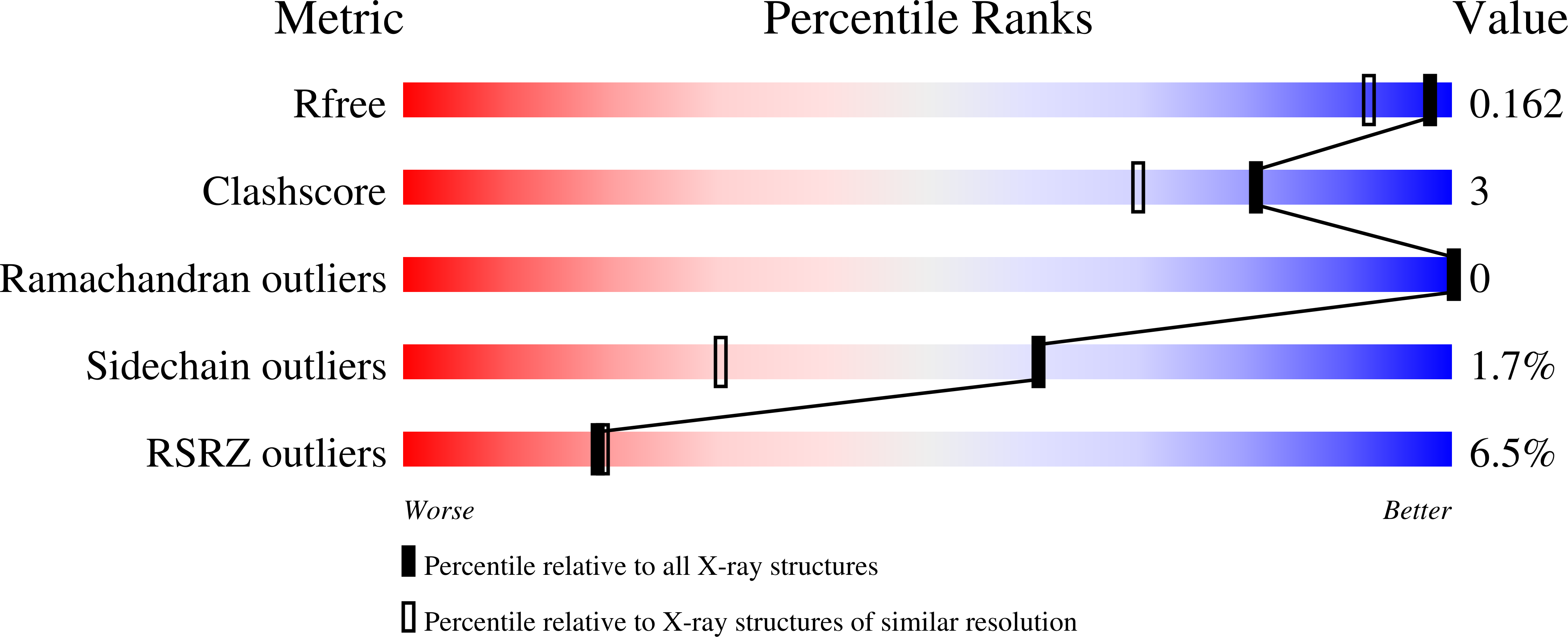
    7QBK, 7QBP

  • PubMed Abstract: 

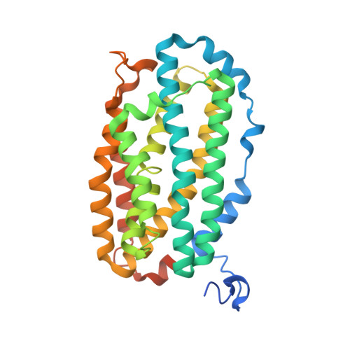
    R2-like ligand-binding oxidase (R2lox) is a ferritin-like protein that harbours a heterodinuclear manganese-iron active site. Although R2lox function is yet to be established, the enzyme binds a fatty acid ligand coordinating the metal centre and catalyses the formation of a tyrosine-valine ether cross-link in the protein scaffold upon O 2 activation. Here, we characterized the ligands copurified with R2lox by mass spectrometry-based metabolomics. Moreover, we present the crystal structures of two new homologs of R2lox, from Saccharopolyspora erythraea and Sulfolobus acidocaldarius, at 1.38 Å and 2.26 Å resolution, respectively, providing the highest resolution structure for R2lox, as well as new insights into putative mechanisms regulating the function of the enzyme.


  • Organizational Affiliation

    Department of Biochemistry and Biophysics, Stockholm University, Stockholm, Sweden.


Macromolecules
Find similar proteins by:  (by identity cutoff)  |  3D Structure
Entity ID: 1
MoleculeChains Sequence LengthOrganismDetailsImage
R2-like ligand binding oxidase330Saccharopolyspora erythraea NRRL 2338Mutation(s): 0 
Gene Names: SACE_0591
EC: 1
UniProt
Find proteins for A4F7B2 (Saccharopolyspora erythraea (strain ATCC 11635 / DSM 40517 / JCM 4748 / NBRC 13426 / NCIMB 8594 / NRRL 2338))
Explore A4F7B2 
Go to UniProtKB:  A4F7B2
Entity Groups  
Sequence Clusters30% Identity50% Identity70% Identity90% Identity95% Identity100% Identity
UniProt GroupA4F7B2
Sequence Annotations
Expand
  • Reference Sequence
Experimental Data & Validation

Experimental Data

  • Method: X-RAY DIFFRACTION
  • Resolution: 1.38 Å
  • R-Value Free: 0.165 
  • R-Value Work: 0.151 
  • R-Value Observed: 0.152 
  • Space Group: P 43 21 2
Unit Cell:
Length ( Å )Angle ( ˚ )
a = 103.173α = 90
b = 103.173β = 90
c = 61.574γ = 90
Software Package:
Software NamePurpose
PHENIXrefinement
XDSdata reduction
XDSdata scaling
PHASERphasing

Structure Validation

View Full Validation Report


Ligand Structure Quality Assessment 


Entry History & Funding Information

Deposition Data


Funding OrganizationLocationGrant Number
European Research Council (ERC)SwedenHIGH-GEAR 724394
Swedish Research CouncilSweden2017-04018
Knut and Alice Wallenberg FoundationSweden2017.0275
Knut and Alice Wallenberg FoundationSweden2019.0436

Revision History  (Full details and data files)

  • Version 1.0: 2022-04-06
    Type: Initial release
  • Version 1.1: 2022-07-06
    Changes: Database references
  • Version 1.2: 2024-01-31
    Changes: Data collection, Refinement description