7PC6

DNA-binding domain of a p53 homolog from the hydrothermal vent annelid Alvinella pompejana


Experimental Data Snapshot

  • Method: X-RAY DIFFRACTION
  • Resolution: 1.92 Å
  • R-Value Free: 0.225 
  • R-Value Work: 0.183 
  • R-Value Observed: 0.186 

wwPDB Validation   3D Report Full Report


This is version 1.1 of the entry. See complete history


Literature

Evolutionary history of the p53 family DNA-binding domain: insights from an Alvinella pompejana homolog.

Zhang, Q.Balourdas, D.I.Baron, B.Senitzki, A.Haran, T.E.Wiman, K.G.Soussi, T.Joerger, A.C.

(2022) Cell Death Dis 13: 214-214

  • DOI: https://doi.org/10.1038/s41419-022-04653-8
  • Primary Citation of Related Structures:  
    7PC6

  • PubMed Abstract: 

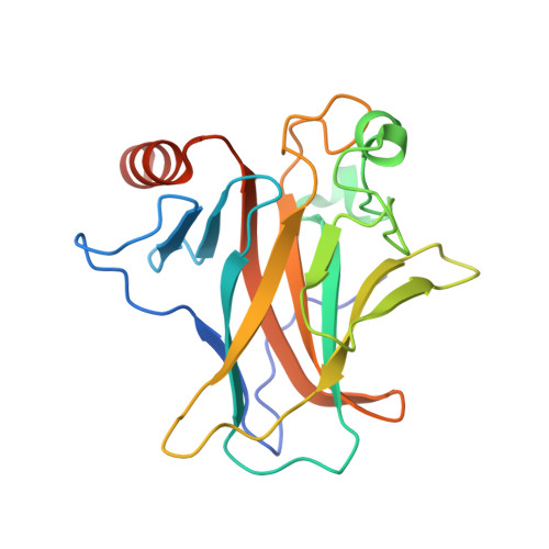
    The extremophile Alvinella pompejana, an annelid worm living on the edge of hydrothermal vents in the Pacific Ocean, is an excellent model system for studying factors that govern protein stability. Low intrinsic stability is a crucial factor for the susceptibility of the transcription factor p53 to inactivating mutations in human cancer. Understanding its molecular basis may facilitate the design of novel therapeutic strategies targeting mutant p53. By analyzing expressed sequence tag (EST) data, we discovered a p53 family gene in A. pompejana. Protein crystallography and biophysical studies showed that it has a p53/p63-like DNA-binding domain (DBD) that is more thermostable than all vertebrate p53 DBDs tested so far, but not as stable as that of human p63. We also identified features associated with its increased thermostability. In addition, the A. pompejana homolog shares DNA-binding properties with human p53 family DBDs, despite its evolutionary distance, consistent with a potential role in maintaining genome integrity. Through extensive structural and phylogenetic analyses, we could further trace key evolutionary events that shaped the structure, stability, and function of the p53 family DBD over time, leading to a potent but vulnerable tumor suppressor in humans.


  • Organizational Affiliation

    Department of Neuroscience, Biomedicum, Karolinska Institutet, Stockholm, Sweden.


Macromolecules
Find similar proteins by:  (by identity cutoff)  |  3D Structure
Entity ID: 1
MoleculeChains Sequence LengthOrganismDetailsImage
DNA-binding domain
A, B, C, D, E
A, B, C, D, E, F, G, H
204Alvinella pompejanaMutation(s): 0 
Entity Groups  
Sequence Clusters30% Identity50% Identity70% Identity90% Identity95% Identity100% Identity
Sequence Annotations
Expand
  • Reference Sequence
Small Molecules
Ligands 2 Unique
IDChains Name / Formula / InChI Key2D Diagram3D Interactions
ZN
Query on ZN

Download Ideal Coordinates CCD File 
I [auth A]
J [auth B]
L [auth C]
M [auth D]
P [auth E]
I [auth A],
J [auth B],
L [auth C],
M [auth D],
P [auth E],
Q [auth F],
R [auth G],
T [auth H]
ZINC ION
Zn
PTFCDOFLOPIGGS-UHFFFAOYSA-N
EDO
Query on EDO

Download Ideal Coordinates CCD File 
K [auth B],
N [auth D],
O [auth D],
S [auth G]
1,2-ETHANEDIOL
C2 H6 O2
LYCAIKOWRPUZTN-UHFFFAOYSA-N
Experimental Data & Validation

Experimental Data

  • Method: X-RAY DIFFRACTION
  • Resolution: 1.92 Å
  • R-Value Free: 0.225 
  • R-Value Work: 0.183 
  • R-Value Observed: 0.186 
  • Space Group: P 1
Unit Cell:
Length ( Å )Angle ( ˚ )
a = 55.9α = 88.074
b = 69.985β = 89.919
c = 114.849γ = 81.512
Software Package:
Software NamePurpose
PHENIXrefinement
XDSdata reduction
Aimlessdata scaling
PHASERphasing

Structure Validation

View Full Validation Report


Entry History & Funding Information

Deposition Data


Funding OrganizationLocationGrant Number
German Research Foundation (DFG)GermanyJO 1473/1-3

Revision History  (Full details and data files)

  • Version 1.0: 2022-03-23
    Type: Initial release
  • Version 1.1: 2024-01-31
    Changes: Data collection, Refinement description