7NDE

Trichoderma parareesei PL7A beta-glucuronan lyase


Experimental Data Snapshot

  • Method: X-RAY DIFFRACTION
  • Resolution: 1.45 Å
  • R-Value Free: 0.174 
  • R-Value Work: 0.155 
  • R-Value Observed: 0.156 

wwPDB Validation   3D Report Full Report


Ligand Structure Quality Assessment 


This is version 1.2 of the entry. See complete history


Literature

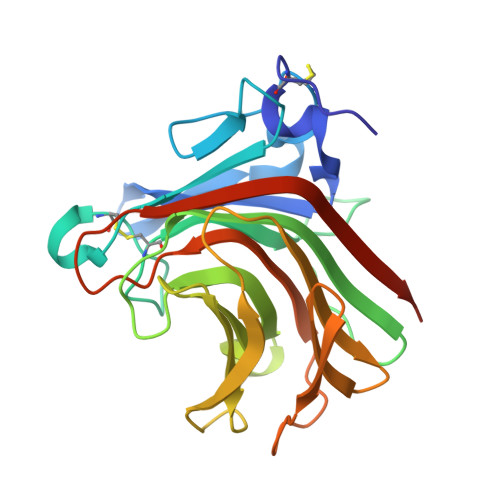
Glucuronan lyases from family PL7 use a Tyr/Tyr syn beta-elimination catalytic mechanism for glucuronan breakdown.

Vuillemin, M.Pilgaard, B.Kiehn, E.Fredslund, F.Welner, D.H.Meyer, A.S.Aachmann, F.L.Wilkens, C.

(2024) Chem Commun (Camb) 60: 440-443

  • DOI: https://doi.org/10.1039/d3cc04256a
  • Primary Citation of Related Structures:  
    7NDE

  • PubMed Abstract: 

    TpPL7A and TpPL7B, members of CAZy family PL7, act as β-glucuronan lyases. TpPL7A diverges by lacking the catalytic histidine, identified as the Brønsted base in PL7 alginate lyases. Our research, including TpPL7A's crystal structure, and mutagenesis studies, reveals a shared syn -β-elimination mechanism with a single tyrosine serving as both base and acid catalyst. This mechanism may extend to subfamily PL7_4 glucuronan lyases.


  • Organizational Affiliation

    Department of Biotechnology and Biomedicine, Technical University of Denmark, Søltofts Plads, Building 224, DK-2800 Kgs. Lyngby, Denmark. cpwk@novonordisk.com.


Macromolecules
Find similar proteins by:  (by identity cutoff)  |  3D Structure
Entity ID: 1
MoleculeChains Sequence LengthOrganismDetailsImage
Glucuronan lyase242Trichoderma parareeseiMutation(s): 0 
Gene Names: A9Z42_0012760
UniProt
Find proteins for A0A2H2ZFD4 (Trichoderma parareesei)
Explore A0A2H2ZFD4 
Go to UniProtKB:  A0A2H2ZFD4
Entity Groups  
Sequence Clusters30% Identity50% Identity70% Identity90% Identity95% Identity100% Identity
UniProt GroupA0A2H2ZFD4
Sequence Annotations
Expand
  • Reference Sequence
Small Molecules
Ligands 2 Unique
IDChains Name / Formula / InChI Key2D Diagram3D Interactions
NAG
Query on NAG

Download Ideal Coordinates CCD File 
B [auth A]2-acetamido-2-deoxy-beta-D-glucopyranose
C8 H15 N O6
OVRNDRQMDRJTHS-FMDGEEDCSA-N
EDO
Query on EDO

Download Ideal Coordinates CCD File 
C [auth A]1,2-ETHANEDIOL
C2 H6 O2
LYCAIKOWRPUZTN-UHFFFAOYSA-N
Experimental Data & Validation

Experimental Data

  • Method: X-RAY DIFFRACTION
  • Resolution: 1.45 Å
  • R-Value Free: 0.174 
  • R-Value Work: 0.155 
  • R-Value Observed: 0.156 
  • Space Group: P 41 21 2
Unit Cell:
Length ( Å )Angle ( ˚ )
a = 69.589α = 90
b = 69.589β = 90
c = 111.66γ = 90
Software Package:
Software NamePurpose
MxCuBEdata collection
XDSdata reduction
XDSdata scaling
Cootmodel building
PHASERphasing
PHENIXrefinement
PHENIXrefinement

Structure Validation

View Full Validation Report


Ligand Structure Quality Assessment 


Entry History 

Deposition Data

Revision History  (Full details and data files)

  • Version 1.0: 2022-03-02
    Type: Initial release
  • Version 1.1: 2024-01-31
    Changes: Data collection, Refinement description
  • Version 1.2: 2024-02-14
    Changes: Database references