7N29

Structure of NAD kinase


Experimental Data Snapshot

  • Method: X-RAY DIFFRACTION
  • Resolution: 2.80 Å
  • R-Value Free: 0.249 
  • R-Value Work: 0.191 
  • R-Value Observed: 0.193 

wwPDB Validation   3D Report Full Report


Ligand Structure Quality Assessment 


This is version 1.1 of the entry. See complete history


Literature

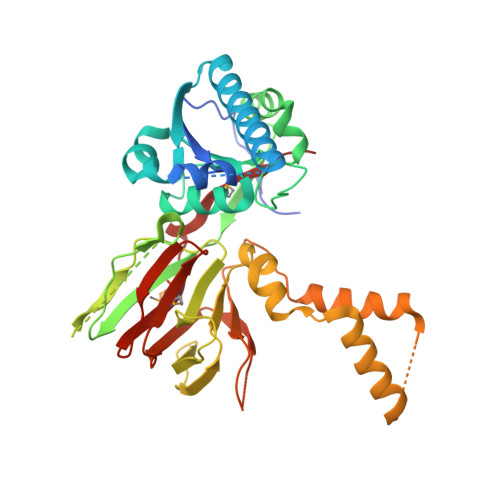
Structure of human NADK2 reveals atypical assembly and regulation of NAD kinases from animal mitochondria.

Du, J.Estrella, M.Solorio-Kirpichyan, K.Jeffrey, P.D.Korennykh, A.

(2022) Proc Natl Acad Sci U S A 119: e2200923119-e2200923119

  • DOI: https://doi.org/10.1073/pnas.2200923119
  • Primary Citation of Related Structures:  
    7N29

  • PubMed Abstract: 

    All kingdoms of life produce essential nicotinamide dinucleotide NADP(H) using NAD kinases (NADKs). A panel of published NADK structures from bacteria, eukaryotic cytosol, and yeast mitochondria revealed similar tetrameric enzymes. Here, we present the 2.8-Å structure of the human mitochondrial kinase NADK2 with a bound substrate, which is an exception to this uniformity, diverging both structurally and biochemically from NADKs. We show that NADK2 harbors a unique tetramer disruptor/dimerization e lement, which is conserved in m itochondrial k inases of a nimals (EMKA) and absent from other NADKs. EMKA stabilizes the NADK2 dimer but prevents further NADK2 oligomerization by blocking the tetramerization interface. This structural change bears functional consequences and alters the activation mechanism of the enzyme. Whereas tetrameric NADKs undergo cooperative activation via oligomerization, NADK2 is a constitutively active noncooperative dimer. Thus, our data point to a unique regulation of NADP(H) synthesis in animal mitochondria achieved via structural adaptation of the NADK2 kinase.


  • Organizational Affiliation

    Department of Molecular Biology, Princeton University, Princeton, NJ 08544.


Macromolecules
Find similar proteins by:  (by identity cutoff)  |  3D Structure
Entity ID: 1
MoleculeChains Sequence LengthOrganismDetailsImage
NAD kinase 2, mitochondrial
A, B, C, D
398Homo sapiensMutation(s): 0 
Gene Names: NADK2C5orf33MNADKNADKD1
EC: 2.7.1.23
UniProt & NIH Common Fund Data Resources
Find proteins for Q4G0N4 (Homo sapiens)
Explore Q4G0N4 
Go to UniProtKB:  Q4G0N4
PHAROS:  Q4G0N4
GTEx:  ENSG00000152620 
Entity Groups  
Sequence Clusters30% Identity50% Identity70% Identity90% Identity95% Identity100% Identity
UniProt GroupQ4G0N4
Sequence Annotations
Expand
  • Reference Sequence
Experimental Data & Validation

Experimental Data

  • Method: X-RAY DIFFRACTION
  • Resolution: 2.80 Å
  • R-Value Free: 0.249 
  • R-Value Work: 0.191 
  • R-Value Observed: 0.193 
  • Space Group: C 2 2 21
Unit Cell:
Length ( Å )Angle ( ˚ )
a = 97.129α = 90
b = 185.115β = 90
c = 188.634γ = 90
Software Package:
Software NamePurpose
PHENIXrefinement
XDSdata reduction
Aimlessdata scaling
SHELXCDphasing
PDB_EXTRACTdata extraction

Structure Validation

View Full Validation Report


Ligand Structure Quality Assessment 


Entry History & Funding Information

Deposition Data


Funding OrganizationLocationGrant Number
National Institutes of Health/National Institute of General Medical Sciences (NIH/NIGMS)United StatesR01GM110161

Revision History  (Full details and data files)

  • Version 1.0: 2022-05-04
    Type: Initial release
  • Version 1.1: 2022-06-29
    Changes: Database references