7N0E

Co-complex of the histidine kinase region of RetS and the dimerization and histidine phosphotransfer domain of GacS


Experimental Data Snapshot

  • Method: X-RAY DIFFRACTION
  • Resolution: 2.30 Å
  • R-Value Free: 0.266 
  • R-Value Work: 0.247 
  • R-Value Observed: 0.249 

wwPDB Validation   3D Report Full Report


This is version 1.2 of the entry. See complete history


Literature

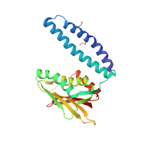
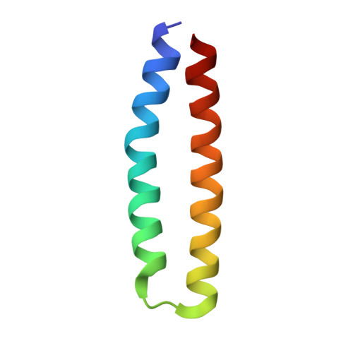
RetS inhibits Pseudomonas aeruginosa biofilm formation by disrupting the canonical histidine kinase dimerization interface of GacS.

Ryan Kaler, K.M.Nix, J.C.Schubot, F.D.

(2021) J Biol Chem 297: 101193-101193

  • DOI: https://doi.org/10.1016/j.jbc.2021.101193
  • Primary Citation of Related Structures:  
    7N0E

  • PubMed Abstract: 

    Bacterial signaling histidine kinases (HKs) have long been postulated to function exclusively through linear signal transduction chains. However, several HKs have recently been shown to form complex multikinase networks (MKNs). The most prominent MKN, involving the enzymes RetS and GacS, controls the switch between the motile and biofilm lifestyles in the pathogenic bacterium Pseudomonas aeruginosa. While GacS promotes biofilm formation, RetS counteracts GacS using three distinct mechanisms. Two are dephosphorylating mechanisms. The third, a direct binding between the RetS and GacS HK regions, blocks GacS autophosphorylation. Focusing on the third mechanism, we determined the crystal structure of a cocomplex between the HK region of RetS and the dimerization and histidine phosphotransfer (DHp) domain of GacS. This is the first reported structure of a complex between two distinct bacterial signaling HKs. In the complex, the canonical HK homodimerization interface is replaced by a strikingly similar heterodimeric interface between RetS and GacS. We further demonstrate that GacS autophosphorylates in trans, thus explaining why the formation of a RetS-GacS complex inhibits GacS autophosphorylation. Using mutational analysis in conjunction with bacterial two-hybrid and biofilm assays, we not only corroborate the biological role of the observed RetS-GacS interactions, but also identify a residue critical for the equilibrium between the RetS-GacS complex and the respective RetS and GacS homodimers. Collectively, our findings suggest that RetS and GacS form a domain-swapped hetero-oligomer during the planktonic growth phase of P. aeruginosa before unknown signals cause its dissociation and a relief of GacS inhibition to promote biofilm formation.


  • Organizational Affiliation

    Department of Biological Sciences, Virginia Polytechnic Institute and State University, Blacksburg, Virginia, USA.


Macromolecules
Find similar proteins by:  (by identity cutoff)  |  3D Structure
Entity ID: 1
MoleculeChains Sequence LengthOrganismDetailsImage
Histidine kinaseA [auth B]225Pseudomonas aeruginosaMutation(s): 0 
Gene Names: GNQ20_09825
EC: 2.7.13.3
UniProt
Find proteins for Q9HUV7 (Pseudomonas aeruginosa (strain ATCC 15692 / DSM 22644 / CIP 104116 / JCM 14847 / LMG 12228 / 1C / PRS 101 / PAO1))
Explore Q9HUV7 
Go to UniProtKB:  Q9HUV7
Entity Groups  
Sequence Clusters30% Identity50% Identity70% Identity90% Identity95% Identity100% Identity
UniProt GroupQ9HUV7
Sequence Annotations
Expand
  • Reference Sequence
Find similar proteins by:  (by identity cutoff)  |  3D Structure
Entity ID: 2
MoleculeChains Sequence LengthOrganismDetailsImage
Histidine kinaseB [auth A]60Pseudomonas aeruginosaMutation(s): 0 
Gene Names: C0044_06730
EC: 2.7.13.3
UniProt
Find proteins for G3XD98 (Pseudomonas aeruginosa (strain ATCC 15692 / DSM 22644 / CIP 104116 / JCM 14847 / LMG 12228 / 1C / PRS 101 / PAO1))
Explore G3XD98 
Go to UniProtKB:  G3XD98
Entity Groups  
Sequence Clusters30% Identity50% Identity70% Identity90% Identity95% Identity100% Identity
UniProt GroupG3XD98
Sequence Annotations
Expand
  • Reference Sequence
Small Molecules
Modified Residues  1 Unique
IDChains TypeFormula2D DiagramParent
MSE
Query on MSE
A [auth B]L-PEPTIDE LINKINGC5 H11 N O2 SeMET
Experimental Data & Validation

Experimental Data

  • Method: X-RAY DIFFRACTION
  • Resolution: 2.30 Å
  • R-Value Free: 0.266 
  • R-Value Work: 0.247 
  • R-Value Observed: 0.249 
  • Space Group: P 31 2 1
Unit Cell:
Length ( Å )Angle ( ˚ )
a = 103.36α = 90
b = 103.36β = 90
c = 61.96γ = 120
Software Package:
Software NamePurpose
PHENIXrefinement
PDB_EXTRACTdata extraction
iMOSFLMdata reduction
Aimlessdata scaling
PHASERphasing

Structure Validation

View Full Validation Report


Entry History & Funding Information

Deposition Data


Funding OrganizationLocationGrant Number
National Institutes of Health/National Institute of General Medical Sciences (NIH/NIGMS)United States--

Revision History  (Full details and data files)

  • Version 1.0: 2022-03-09
    Type: Initial release
  • Version 1.1: 2023-10-18
    Changes: Data collection, Refinement description
  • Version 1.2: 2023-11-15
    Changes: Data collection