7LK4

Crystal structure of BAK L100A in complex with activating antibody fragments


Experimental Data Snapshot

  • Method: X-RAY DIFFRACTION
  • Resolution: 3.10 Å
  • R-Value Free: 0.262 
  • R-Value Work: 0.214 
  • R-Value Observed: 0.216 

wwPDB Validation   3D Report Full Report


This is version 1.3 of the entry. See complete history


Literature

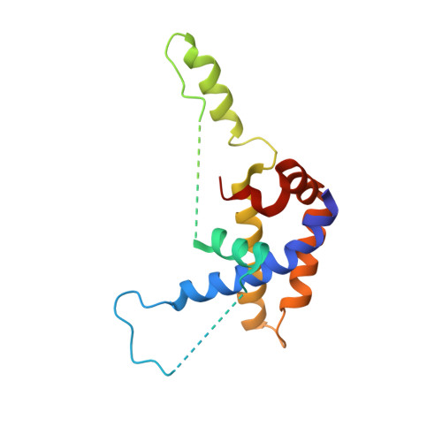
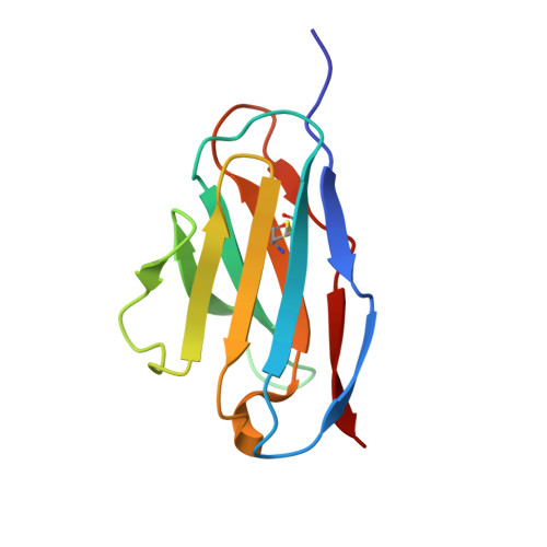
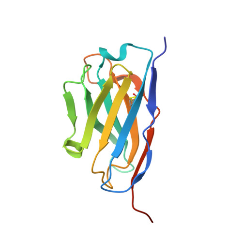
Structure of the BAK-activating antibody 7D10 bound to BAK reveals an unexpected role for the alpha 1-alpha 2 loop in BAK activation.

Robin, A.Y.Miller, M.S.Iyer, S.Shi, M.X.Wardak, A.Z.Lio, D.Smith, N.A.Smith, B.J.Birkinshaw, R.W.Czabotar, P.E.Kluck, R.M.Colman, P.M.

(2022) Cell Death Differ 29: 1757-1768

  • DOI: https://doi.org/10.1038/s41418-022-00961-w
  • Primary Citation of Related Structures:  
    7LK4

  • PubMed Abstract: 

    Pro-apoptotic BAK and BAX are activated by BH3-only proteins to permeabilise the outer mitochondrial membrane. The antibody 7D10 also activates BAK on mitochondria and its epitope has previously been mapped to BAK residues in the loop connecting helices α1 and α2 of BAK. A crystal structure of the complex between the Fv fragment of 7D10 and the BAK mutant L100A suggests a possible mechanism of activation involving the α1-α2 loop residue M60. M60 mutants of BAK have reduced stability and elevated sensitivity to activation by BID, illustrating that M60, through its contacts with residues in helices α1, α5 and α6, is a linchpin stabilising the inert, monomeric structure of BAK. Our data demonstrate that BAK's α1-α2 loop is not a passive covalent connector between secondary structure elements, but a direct restraint on BAK's activation.


  • Organizational Affiliation

    Structural Biology Division, The Walter and Eliza Hall Institute of Medical Research, 1G Royal Parade, Parkville, Melbourne, VIC, 3052, Australia.


Macromolecules
Find similar proteins by:  (by identity cutoff)  |  3D Structure
Entity ID: 1
MoleculeChains Sequence LengthOrganismDetailsImage
Bcl-2 homologous antagonist/killerA [auth P],
B [auth Q],
C [auth R],
D [auth S]
164Homo sapiensMutation(s): 2 
Gene Names: BAK1BAKBCL2L7CDN1
UniProt & NIH Common Fund Data Resources
Find proteins for Q16611 (Homo sapiens)
Explore Q16611 
Go to UniProtKB:  Q16611
PHAROS:  Q16611
GTEx:  ENSG00000030110 
Entity Groups  
Sequence Clusters30% Identity50% Identity70% Identity90% Identity95% Identity100% Identity
UniProt GroupQ16611
Sequence Annotations
Expand
  • Reference Sequence
Find similar proteins by:  (by identity cutoff)  |  3D Structure
Entity ID: 2
MoleculeChains Sequence LengthOrganismDetailsImage
7D10 antibody VL fragmentE [auth A],
G [auth C],
I [auth E],
K [auth G]
109Rattus norvegicusMutation(s): 0 
Entity Groups  
Sequence Clusters30% Identity50% Identity70% Identity90% Identity95% Identity100% Identity
Sequence Annotations
Expand
  • Reference Sequence
Find similar proteins by:  (by identity cutoff)  |  3D Structure
Entity ID: 3
MoleculeChains Sequence LengthOrganismDetailsImage
7D10 antibody VH fragmentF [auth B],
H [auth D],
J [auth F],
L [auth H]
129Rattus norvegicusMutation(s): 0 
Entity Groups  
Sequence Clusters30% Identity50% Identity70% Identity90% Identity95% Identity100% Identity
Sequence Annotations
Expand
  • Reference Sequence
Experimental Data & Validation

Experimental Data

  • Method: X-RAY DIFFRACTION
  • Resolution: 3.10 Å
  • R-Value Free: 0.262 
  • R-Value Work: 0.214 
  • R-Value Observed: 0.216 
  • Space Group: P 1 21 1
Unit Cell:
Length ( Å )Angle ( ˚ )
a = 57.17α = 90
b = 271.8β = 91.79
c = 74.04γ = 90
Software Package:
Software NamePurpose
PHENIXrefinement
XDSdata reduction
PHASERphasing
PDB_EXTRACTdata extraction
XDSdata scaling

Structure Validation

View Full Validation Report


Entry History 

Deposition Data

Revision History  (Full details and data files)

  • Version 1.0: 2022-02-09
    Type: Initial release
  • Version 1.1: 2022-08-24
    Changes: Database references
  • Version 1.2: 2022-09-14
    Changes: Database references
  • Version 1.3: 2023-10-18
    Changes: Data collection, Refinement description