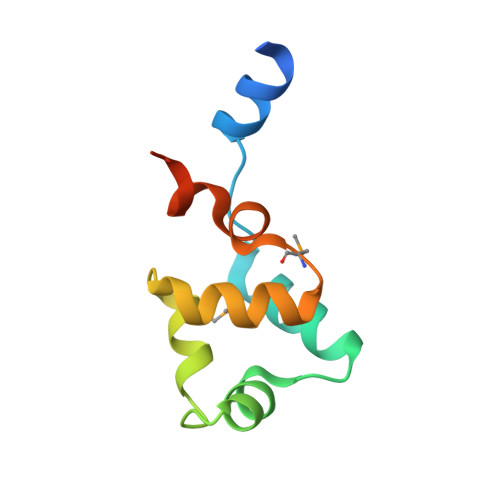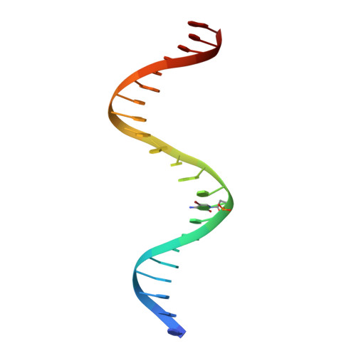
Xenogeneic nucleoid-associated EnrR thwarts H-NS silencing of bacterial virulence with unique DNA binding.
Ma, R., Liu, Y., Gan, J., Qiao, H., Ma, J., Zhang, Y., Bu, Y., Shao, S., Zhang, Y., Wang, Q.(2022) Nucleic Acids Res 50: 3777-3798
- PubMed: 35325196
- DOI: https://doi.org/10.1093/nar/gkac180
- Primary Citation of Related Structures:
7F9H, 7F9I - PubMed Abstract:
Type III and type VI secretion systems (T3/T6SS) are encoded in horizontally acquired genomic islands (GIs) that play crucial roles in evolution and virulence in bacterial pathogens. T3/T6SS expression is subjected to tight control by the host xenogeneic silencer H-NS, but how this mechanism is counteracted remains to be illuminated. Here, we report that xenogeneic nucleoid-associated protein EnrR encoded in a GI is essential for virulence in pathogenic bacteria Edwardsiella and Salmonella. We showed that EnrR plays critical roles in T3/T6SS expression in these bacteria. Various biochemical and genetic analyses demonstrated that EnrR binds and derepresses the promoter of esrB, the critical regulator of T3/T6SS, to promote their expression by competing with H-NS. Additionally, EnrR targets AT-rich regions, globally modulates the expression of ∼363 genes and is involved in various cellular processes. Crystal structures of EnrR in complex with a specific AT-rich palindromic DNA revealed a new DNA-binding mode that involves conserved HTH-mediated interactions with the major groove and contacts of its N-terminal extension to the minor groove in the symmetry-related duplex. Collectively, these data demonstrate that EnrR is a virulence activator that can antagonize H-NS, highlighting a unique mechanism by which bacterial xenogeneic regulators recognize and regulate foreign DNA.
Organizational Affiliation:
State Key Laboratory of Bioreactor Engineering, East China University of Science and Technology, Shanghai 200237, China.