7EZG

The structure of the human METTL6 enzyme in complex with SAH


Experimental Data Snapshot

  • Method: X-RAY DIFFRACTION
  • Resolution: 1.90 Å
  • R-Value Free: 0.201 
  • R-Value Work: 0.179 
  • R-Value Observed: 0.180 

wwPDB Validation   3D Report Full Report


Ligand Structure Quality Assessment 


This is version 1.1 of the entry. See complete history


Literature

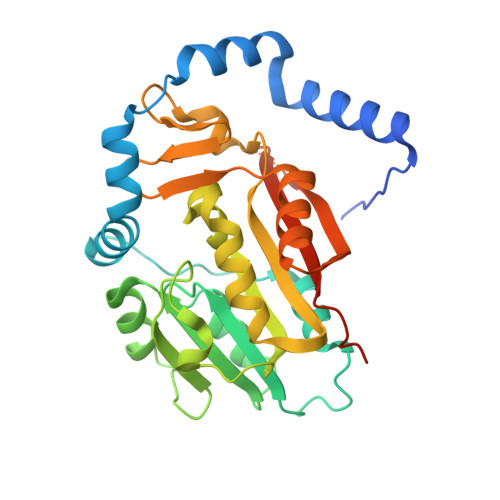
Crystal structure of human METTL6, the m 3 C methyltransferase.

Chen, R.Zhou, J.Liu, L.Mao, X.L.Zhou, X.Xie, W.

(2021) Commun Biol 4: 1361-1361

  • DOI: https://doi.org/10.1038/s42003-021-02890-9
  • Primary Citation of Related Structures:  
    7EZG

  • PubMed Abstract: 

    In tRNA, the epigenetic m 3 C modification at position 32 in the anticodon loop is highly conserved in eukaryotes, which maintains the folding and basepairing functions of the anticodon. However, the responsible enzymes METTL2 and METTL6 were identified only in recent years. The loss of human METTL6 (hMETTL6) affects the translational process and proteostasis in cells, while in mESCs cells, it leads to defective pluripotency potential. Despite its important functions, the catalytic mechanism of the C32 methylation by this enzyme is poorly understood. Here we present the 1.9 Å high-resolution crystal structure of hMETTL6 bound by SAH. The key residues interacting with the ligand were identified and their roles were confirmed by ITC. We generated a docking model for the hMETTL6-SAH-CMP ternary complex. Interestingly, the CMP molecule binds into a cavity in a positive patch with the base ring pointing to the inside, suggesting a flipped-base mechanism for methylation. We further generated a model for the quaternary complex with tRNA Ser as a component, which reasonably explained the biochemical behaviors of hMETTL6. Taken together, our crystallographic and biochemical studies provide important insight into the molecular recognition mechanism by METTL6 and may aid in the METTL-based rational drug design in the future.


  • Organizational Affiliation

    MOE Key Laboratory of Gene Function and Regulation, State Key Laboratory for Biocontrol, School of Life Sciences, The Sun Yat-Sen University, Guangzhou, Guangdong, 510006, People's Republic of China.


Macromolecules
Find similar proteins by:  (by identity cutoff)  |  3D Structure
Entity ID: 1
MoleculeChains Sequence LengthOrganismDetailsImage
tRNA N(3)-methylcytidine methyltransferase METTL6305Homo sapiensMutation(s): 0 
Gene Names: METTL6
EC: 2.1.1
UniProt & NIH Common Fund Data Resources
Find proteins for Q8TCB7 (Homo sapiens)
Explore Q8TCB7 
Go to UniProtKB:  Q8TCB7
PHAROS:  Q8TCB7
GTEx:  ENSG00000206562 
Entity Groups  
Sequence Clusters30% Identity50% Identity70% Identity90% Identity95% Identity100% Identity
UniProt GroupQ8TCB7
Sequence Annotations
Expand
  • Reference Sequence
Small Molecules
Ligands 1 Unique
IDChains Name / Formula / InChI Key2D Diagram3D Interactions
SAH (Subject of Investigation/LOI)
Query on SAH

Download Ideal Coordinates CCD File 
B [auth A]S-ADENOSYL-L-HOMOCYSTEINE
C14 H20 N6 O5 S
ZJUKTBDSGOFHSH-WFMPWKQPSA-N
Experimental Data & Validation

Experimental Data

  • Method: X-RAY DIFFRACTION
  • Resolution: 1.90 Å
  • R-Value Free: 0.201 
  • R-Value Work: 0.179 
  • R-Value Observed: 0.180 
  • Space Group: P 2 3
Unit Cell:
Length ( Å )Angle ( ˚ )
a = 110.264α = 90
b = 110.264β = 90
c = 110.264γ = 90
Software Package:
Software NamePurpose
PHENIXrefinement
HKL-2000data reduction
HKL-2000data scaling
PHASERphasing

Structure Validation

View Full Validation Report


Ligand Structure Quality Assessment 


Entry History & Funding Information

Deposition Data


Funding OrganizationLocationGrant Number
National Natural Science Foundation of China (NSFC)China31870782

Revision History  (Full details and data files)

  • Version 1.0: 2022-01-12
    Type: Initial release
  • Version 1.1: 2023-11-29
    Changes: Data collection, Refinement description